ASH 2011 Bystander vaccine poster (PDF)
File information
This PDF 1.4 document has been generated by Illustrator / Adobe PDF library 6.66, and has been sent on pdf-archive.com on 08/12/2011 at 20:53, from IP address 206.81.x.x.
The current document download page has been viewed 1777 times.
File size: 18.16 MB (1 page).
Privacy: public file
File preview
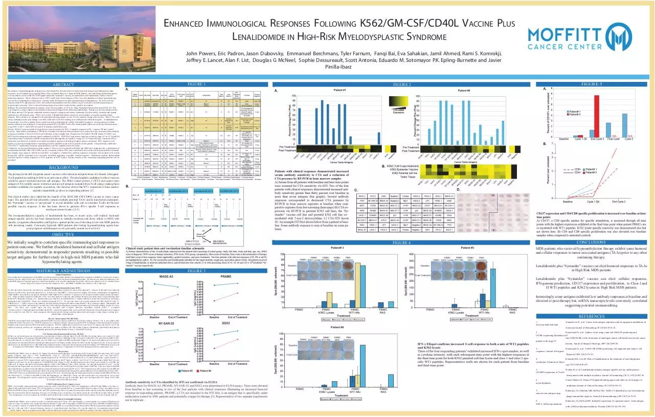
ENHANCED IMMUNOLOGICAL RESPONSES FOLLOWING K562/GM-CSF/CD40L VACCINE PLUS
LENALIDOMIDE IN HIGH-RISK MYELODYSPLASTIC SYNDROME
John Powers, Eric Padron, Jason Dubovsky, Emmanuel Berchmans, Tyler Farnum, Fanqi Bai, Eva Sahakian, Jamil Ahmed, Rami S. Komrokji,
Jeffrey E. Lancet, Alan F. List, Douglas G McNeel, Sophie Dessureault, Scott Antonia, Eduardo M. Sotomayor P.K. Epling-Burnette and Javier
Pinilla-Ibarz
ABSTRACT
FIGURE 1
FIGURE 5
FIGURE 2
Introduction: Immunotherapeutic strategies have been limited by the lack of known tumor restricted antigens and inadequate immune
responses. In our Institution, an ongoing Phase I dose escalation study in 11 high risk MDS patients, who failed hypo-methylating agents,
were enrolled in a K562-GMCSF-CD40 ligand transfected “bystander” vaccine, in combination with lenalidomide, and have shown
promising responses. Wilm’s Tumor protein 1 (WT1) and Cancer Testis Antigens (CTAs) are solid candidates for future immunotherapy
and vaccines containing these antigens are currently under development in solid and hematological malignancies. The K562 cell-line
expresses both WT1 and numerous CTAs, the resultant immunization with this cellular vaccine is attractive for both immunological
response and assessment. Here we describe immunological correlative studies before and after vaccination.
Methods: We monitored the humoral response, of the four responders, to CTAs by High Throughput Immunoblot assay (HTI), 29 CTAs
were bound in a concise pattern to nitrocellulose filters and developed in the following methodology. Patient sera were incubated with the
HTI filters and any CTA specific antibodies bound to respective antigens and reactive antibodies were detected by secondary anti-human
antibodies in a colorimetric assay. Filters were read by 6 blinded individuals, spots were scored positive or negative against control
antigens. These results were compared to the paired post-therapy sample a score of 6 was maximal change of pre to post. Those CTAs with
strong antibody presence post-therapy were then confirmed via ELISA resulting in an antibody titer reflected as ug/mL. A standardization
of human IgG, as well as a healthy donor control was used to determine the validity of identified responses. An assessment of cellular
immune responses was performed via interferon gamma (IFNγ) ELISPOT and a flow based peptide and lysate specific proliferation assay,
using CD8+, CD137+ and CSFE, towards WT1 peptides and K562 lysates.
Results: Of the 15 patients enrolled 4 had relevant clinical responses by IWG, 2 complete responses (CR), 1 marrow CR and 1 partial
response. Upon initial examination of HTI filters, 4 patients who showed clinical response were observed to have increased serum antibody
responses to multiple CTAs (10 of 29) at end of therapy or last evaluable date. Response to NY-SAR-35, MAGE family, NY-ESO1 and
SSX2 had the strongest post-therapy signal confirmed via ELISA. RT-PCR of bone marrow aspirates revealed a range of 5 to 9 CTA mRNA
transcripts present in responsive patients at baseline which subsequently decreased to a range of 1 to 5 at post-therapy. Increased (IFNγ)
production was observed via elispot in an initial screening of a patient with clinical response at post-vaccination. IFNγ responsive cell
number was increased independent to macrophage loaded with K562 lysate or WT1 peptides in this patient. This patient also exhibited a
CD8+CD137+ proliferative response upon incubation with WT1 peptides and K562 lysate.
Conclusion: An increased humoral response against CTAs was seen in patients with high risk MDS after treatment with a combination of
lenalidomide and K562-GM-CSF-CD40L vaccine. A majority of these CTAs were expressed by the vaccine and the patients before treatment.
Interestingly, some antigens, MAGE-A4 and SSX1 as examples, exhibited low antibody responses at baseline and elevated levels at post
therapy but, mRNA transcripts were conversely correlated suggesting a possible immune response. Preliminary analysis demonstrated
increased specific cellular responses to WT1 peptides or K562 lysates. Further analysis of the remaining responding patients will be
presented.
BACKGROUND
The primary down-fall of peptide cancer vaccines is the induction and persistence of a biased, tolerogenic
T-cell population resulting in little to no anti-cancer effect. The ideal peptide candidates for these vaccines
would be cancer restricted and non-self antigens. The Wilm’s tumor protein 1 (WT1) and cancer testis
antigens (CTA) exhibit cancer restriction and are self antigens excluded from T-cell anergy making them
excellent candidates for peptide vaccination. Our data has shown that WT1 expression in bone marrow
aspirates sequentially go down in responding patients. [1]
Past clinical studies have identified the benefit of the K562/GM-CSF/CD40L vaccine in many cancer
types. The parental cell line inherently contains multiple potential TAA’s and its transfected counterpart,
the “bystander” vaccine, is “pre-primed” to recruit dendritic cells and co-stimulate T-cells for the best
possible vaccine response. It also has been shown to generate IFN-γ specific T-cell responses to
autologous tumor lysates. [2-3].
The immunomodulatory capacity of lenalidomide has been, in recent years, well studied. Increased
antigen-specific activity has been demonstrated in multiple myeloma and direct effects in MDS with
specific cytogenetic abnormalities (del5q) have opened up the use of this drug in low-risk MDS patients
with promising results. Conversely high-risk MDS patients after failing hypomethylating agents have
poor prognosis and therapies revolve primarily around survival [4-6].
OBJECTIVE
We initially sought to correlate specific immunological responses to
patient outcome. We further elucidated humoral and cellular antigen
sensitivity demonstrated in responder patients resulting in possible
target antigens for further study in high-risk MDS patients who fail
hypomethylating agents.
MATERIALS AND METHODS
Subject Populations
Sera, peripheral blood mononuclear cells (PBMCs) and bone marrow aspirates (BMs) were obtained from 16 patients with MDS (13 males and 3 females,
mean age 50 years, range 64-86 years). Of these subjects, all patients received prior hypomethylating agents. All subjects gave written institutional review
board (IRB)-approved informed consent for their blood products to be used for immunological research. Blood was collected at H. Lee Moffitt Cancer
Center (Tampa, FL), plasma were stored in aliquots at -80°C , and PBMCs and BMs were stored at -140°C until used.
High-Throughput Immunoblot assay (HTI)
We have previously reported the construction of a panel of lambda phage encoding 29 cancer-testis antigens[7]. Analysis of this panel was conducted
similarly to what we have previously reported [7, 8]. In brief, XL-1 blue MRF E. coli were grown overnight, collected by centrifugation, resuspended in
10mM MgSO4, and poured in top agarose (LB broth/10mM MgSO4/0.2% maltose/0.7% agarose) over LB agar in Omniwell plates (Nunc, Rochester,
NY). Phage encoding individual (9,000 pfu) CTA’s were then spotted in replicates onto multiple bacterial agar lawns using a liquid handling robot
(Biomek FX, Beckman, Fullerton, CA). Spotted plates were allowed to sit undisturbed for 15 minutes and then overlaid with nitrocellulose membranes
impregnated with 10mM IPTG. Plates were incubated overnight at 37°C. The next day, filters were washed, blocked with TBS (50mM Tris pH 7.2,
100mM NaCL) + 1%BSA (bovine serum albumin), and then probed overnight with human serum diluted 1:100 in blocking solution. Subsequently, the
membranes were washed, and human IgG was detected with an alkaline phosphatase-conjugated anti-human IgG detection antibody (Sigma, St. Louis,
MO).
The filters were washed again and then developed with 0.3mg/ml nitro-blue tetrazolium chloride (NBT) + 0.15mg/ml
5-bromo-4-chloro-3’-indolyphosphate p-toluidine salt (BCIP). After development, filters were washed with deionized water and immunoreactive plaques
were recorded for each filter by visual comparison with internal positive (phage encoding human IgG) and negative (empty phage encoding betagalactosidase control plaques.
Reverse Transcriptase-PCR (RT-PCR)
Total RNA was prepared from centrifugally pelleted BM (RNeasy mini columns and RNAse free DNAse, Qiagen, Valencia, CA) or was commercially
obtained (BioChain, Hayward, CA). RT-PCR reactions were conducted using the Qiagen one-step RT-PCR kit (Qiagen) with transcript-specific primers
(Suppled upon request) and total RNA from BM as templates. RT-PCR amplification reactions were resolved on 2% agarose gels and the size of the
amplified transcript confirmed by comparison with DNA size markers (GelPilot 1Kb Plus Ladder, Qiagen). Densitometry determined and heatmaps
subsequently generated by Heatmap Builder Version 1.1 (Stanford University)
CTA Enzyme-linked Immunosorbent Assay (ELISA)
ELISA was preformed using sera obtained from non-heparinized blood collection tubes. 96 well EIA/RIA flat bottom plates were coated O/N with
Recombinant Human CTA antigens (PRAME H00023532-P01, MAGE-A3 H00004102-P01, NY-SAR-35 H00158521-P01 and SSX2 H00006757-P01
Novus Biologicals) at 10ug/mL with 100uL per well. Plates were washed with PBS and blocked. Diluted (1:25) and serially diluted sera was used in
conjunction with a standard of Purified Human IgG antibody (AG711 Millipore) serially diluted 8 to 0.0625 ug/mL. Incubate O/N at 4C. Sera was removed
and the plate was incubated with a 1:1000 diluted horseradish peroxidase conjugated, anti-human IgG detection antibody (555788 BD Biosciences) used
in conjunction with SureBlue TMB microwell peroxidase substrate (1-component) (52-00-01 KPL Kirkegaard & Perry Laboratories, Inc.)
Elispot assay
200,000 Whole PBMCs were co-cultured, in U-bottom 96 well plate with media alone, 5x freeze/thaw K562 lysate (30,000 cells equivalent), WT1 mixed
peptide (20ug/mL each Class I -WT1, RMFPNAPYL, Class II WT1331L, PGCNKRYFKLSHLQMHSRKHTG, and Class II-WT1427L,
RSDELVRHHNMHQRNMTKL), (-) control peptide (60ug/mL RAS, TEYKLVVVGAPGVGKSALTI) or (+) control (PMA/Iono), 1ug/mL Phorbol 12myristate 13-acetate (P1585 Sigma-Aldrich) and 0.1ug/mL Ionomycin (I9657 Sigma- Aldrich) for O/N at 37C. Elispot plates (MAIP #S4510) were
prepared on the same day by pre-soaking in 35%EtOH/H2O then rinsing 6x with distilled autoclaved water. Plates were washed 3x with PBS and
100uL/well of 5ug/mL anti-human IFN-gamma antibody (3420-3-1000 Mabtech Clone 1-D1K, purified ) was add and incubated O/N at 4C. On the
following day the Elispot plate was rinsed 3X with RPMI and block for 3hrs. The Elispot Plate was then rinsed with RPMI. Cells were rinsed with RPMI
and resuspended in co-culture media and added to prepared Elispot plate and incubated O/N at 37C. The Elispot plate was then washed 6X with PBS and
0.05% Tween-20 (P7949 Sigma-Aldrich) and 100uL/well of 1ug/mL biotinylated Anti-Human IFN-gamma antibody (3420-6-1000 Mabtech Clone 7-B6-1
) in PBS/0.05%Tween was add and the plate was incubated for O/N at 4C. The following day the Elispot plate was then washed 6X with PBS/0.05%Tween
and 100uL/well of (1:1000) Avidin-HRP (554058 BD Pharmingen ) in PBS/0.05% Tween was add and the plate was incubated for 1hr at room
temperature. The Elispot plate was then washed 6X with PBS/0.05%Tween and 3X with PBS. Spots were developed with AEC buffer (551951 BD
Bioscience) and read on AID spot reader.
CFSE Proliferation Flow Cytometry Assay
PBMCs were initially stained, following product protocols, with 0.2 uM CFDA SE (V12883 Life technologies). 200,000 Whole PBMCs were then cocultured with media alone, K562 lysate, WT1 mixed peptide, (-) control peptide RAS or (+) control PMA/Iono for a total of 5 days. All cultures were in
RPMI/10%FBS/Pen-Strep. Separation of CD4+ and CD8+ proliferating cells were conducted using fluorochrome-labeled monoclonal antibodies (mAbs;
anti-CD3 and anti-CD8, Becton Dickinson, San Jose, CA and eBiosciences, San Diego, CA) and the vitality dye LIVE/DEAD Fixable Yellow (L34959
Life technologies). Data was acquired on an LSRII cytometer (Beckman Coulter), and analyzed with FlowJo software (Tree Star, Ashland, OR). [9]
CD137 Flow Cytometry Immunophenotyping
200,000 Whole PBMCs were co-cultured, as above, with media alone, K562 lysate, WT1 mixed peptide, (-) control peptide or (+) control, (PMA/Iono) for
a total of 4 days. All cultures were washed and cultured another 12 hours with fresh peptides and PMA/iono respectively in RPMI/10%FBS/Pen-Strep.
Flow cytometric analysis of peripheral blood mononuclear cells (PBMCs) was performed using fluorochrome-labeled monoclonal antibodies (mAbs;
anti-CD3, -CD8, and -CD137, Becton Dickinson, San Jose, CA and eBiosciences, San Diego, CA) and the vitality dye 4',6-diamidino-2-phenylindole
(DAPI, Sigma). Data was acquired on an LSRII cytometer (Beckman Coulter), and analyzed with FlowJo software (Tree Star, Ashland, OR). [10]
CONCLUSIONS
FIGURE 4
MDS patients who recieved hypomethylation therapy exhibit some humoral
and cellular responses to tumor associated antigens (TAAs) prior to any other
continuing therapy.
FIGURE 3
Lenalidomide plus “bystander” vaccine can elicit humoral responses to TAAs
in High Risk MDS patients
Lenalidomide plus “bystander” vaccine can elicit cellular responses,
IFN-gamma production, CD137 expression and proliferation, to Class I and
II WT1 peptides and K562 lysates in High Risk MDS patients
Interestingly some antigens exhibited low antibody responses at baseline and
elevated at post-therapy but, mRNA transcripts levels conversely correlated
suggesting potential immunoediting
REFERENCES
1.
acute myeloid leukemia.
Atanackovic D, et al. Cancer testis antigen expression and its epigenetic modulation in
American Journal of Hematology 2011;86(11)918-22.
2.
Dessureault S, et al. A phase-I trial using a univeral GM-CSF-producing and
CD40L-expressing Bystander cell
line (GM.CD40L) in the formation of autologous tumor cell-based vaccines for cancer
patients with stage IV
disease. Annals of Surgical Oncology 2007;14(2)869-84.
3.
Dessureault S, et al. A GM-CSF/CD40L producing cell augments anti-tumor t cell
responses. Journal of Surgical
Research 2005;125(2)173-81.
4.
Komrokji RS, List AF. Role of lenalidomide in the treatment of myelodysplastic
syndromes. Seminars in Oncol
ogy 2011;38(5)648-657.
5.
Neuber B, et al. Lenalidomide enhances antigen-specific activity and decreases
CD45RA expression of T cells
from patients with multiple myeloma. Journal of Immunology 2011; 187(2)1047-56.
6.
myelodysplastic
Garcia-Manero G, Fenaux P. Hypomethylating agents and other novel strategies in
7.
cancer-testis antigen using
Dubovsky JA, Albertini MR, McNeel DG. MAD-CT identified as a novel melanoma
8.
SSX-2, following treatment
Dubovsky JA, McNeel DG. Inducible expression of a prostate cancer -testis antigen,
syndromes.Journal of Clinical Oncology 2011;29(5)516-23.
phage immunoblot analysis. Journal of Immunotherapy 2007;30(7);675-83.
with a DNA methylation inhibitor. Prostate 2007;67(16)1781-90.
Download ASH 2011 Bystander vaccine poster
ASH 2011 Bystander vaccine poster.pdf (PDF, 18.16 MB)
Download PDF
Share this file on social networks
Link to this page
Permanent link
Use the permanent link to the download page to share your document on Facebook, Twitter, LinkedIn, or directly with a contact by e-Mail, Messenger, Whatsapp, Line..
Short link
Use the short link to share your document on Twitter or by text message (SMS)
HTML Code
Copy the following HTML code to share your document on a Website or Blog
QR Code to this page

This file has been shared publicly by a user of PDF Archive.
Document ID: 0000035679.