2013 lamour j.nanosci.lett (PDF)
File information
This PDF 1.3 document has been sent on pdf-archive.com on 16/05/2012 at 20:47, from IP address 137.82.x.x.
The current document download page has been viewed 1687 times.
File size: 1.9 MB (4 pages).
Privacy: public file


File preview
JOURNAL OF NANOSCIENCE LETTERS
Tuning surface energy at the nanometer scale:
A new step towards controlling neuronal differentiation?
Guillaume Lamoura,b, Sylvie Souèsd, Emilie Collarta, Stéphane Colline, Nathalie Bardoue, Ahmed Hamraouia,c,*
a
Université Paris Descartes, UFR Biomédicale, 45 rue des Saints-Pères, 75270 Paris Cedex 06, France
b
Present address: University of British Columbia, Vancouver, B. C., Canada V6T1Z4
c
CEA-Saclay, Service de Physique et Chimie des Surfaces et Interfaces, 91191 Gif-sur- Yvette, France
d
Université Paris Descartes, Génétique Moléculaire et Défense antivirale, FRE3235, 45 rue des Saints-Pères, 75006 Paris, France
e
CNRS, Lab Photon & Nanostruct LPN, F-91460 Marcoussis, France
*
Author for correspondence: Ahmed Hamraoui, email: ahmed.hamraoui@cea.fr
Received 4 Feb 2012; Accepted 16 Apr 2012; Available Online 18 Apr 2012
Abstract
In this paper, we study the impact of the spatial distribution of surface energy on the ability of PC12 cells to extend neurites, which are
projections from the cell body characteristic of differentiating neurons. PC12 cells are a well-known model for studying the mechanisms of
neuronal differentiation that is typically induced by a treatment with a soluble factor, the “nerve growth factor” (NGF). In recent papers, we
showed that PC12 differentiation could be triggered through cell-surface interactions only, providing cell culture substrates displayed nanoscale
heterogeneities. Here, we use patterned surfaces to finely tune the magnitude of the surface energy gradients and total surface energy and
compare their respective propensity to trigger PC12 neurite outgrowth. Glass surfaces were crafted with nanometer-sized gold pillars as culture
substrates. Our data indicate that PC12 cells are sensitive to the respective amounts of gold and glass, and show a better ability to differentiate on
surfaces containing the highest proportion of gold. Precise thresholds regarding the spatial range and the magnitude of the surface energy
gradients critical to the cell response could not be assigned here. However, our results pave the way for new experiments where surface
functionalization combined to the surface patterning strategies used here should allow us to quantify parameters that, once known, would be of
great value to the design of biomaterials for tissue engineering applications.
Keywords: PC12 cells; Neurite outgrowth; Nanostructure; Nanopillars; Surface energy; Patterned surface
1. Introduction
The extraordinary abilities of the nervous system are
based on its functional unit, the neuronal cell. The human
brain, for example, has a number of neurons close to one
hundred billion, and one of these neurons can connect up to
one hundred thousand other neurons. In terms of
combinatorial possibilities, this corresponds to a huge number
of potential connections that defies the imagination. Of
course, these connections, known as synapses, cannot be
determined randomly otherwise the system could no longer
ensure its duties properly. Hence one aspect of the complexity
displayed by the nervous system is the growth of ordered and
oriented nervous fibers during embryogenesis.
The
physicochemical mechanisms responsible for the resulting
complex structure that constitutes the brain or the spinal cord
are far from being fully elucidated. However the impact of
chemical, spatial and mechanical cues of cell culture
substrates have been demonstrated throughout several studies.
In particular, surface adhesion parameters, that control cell
functions together with the genetic program of the cell, have
become better known over the past few years [1-4]. More
generally, a better understanding of cell-substrate interactions
is necessary towards substantial progress in designing
biomaterials consistent with tissue engineering strategies to
promote functional repair of damaged axons.
Here we focus on the surface energy parameter, and
in particular, on the influence of the spatial distribution of the
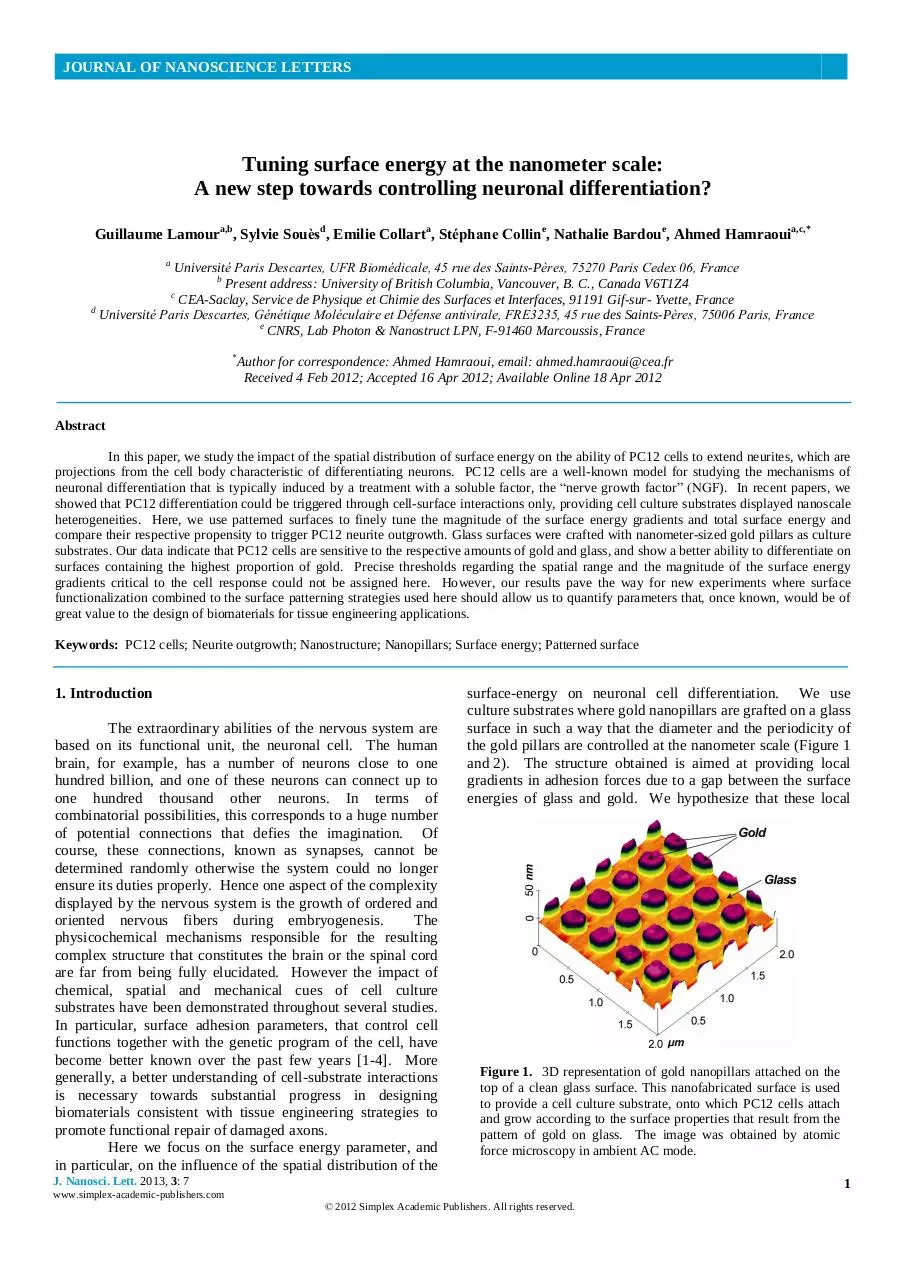
surface-energy on neuronal cell differentiation. We use
culture substrates where gold nanopillars are grafted on a glass
surface in such a way that the diameter and the periodicity of
the gold pillars are controlled at the nanometer scale (Figure 1
and 2). The structure obtained is aimed at providing local
gradients in adhesion forces due to a gap between the surface
energies of glass and gold. We hypothesize that these local
Figure 1. 3D representation of gold nanopillars attached on the
top of a clean glass surface. This nanofabricated surface is used
to provide a cell culture substrate, onto which PC12 cells attach
and grow according to the surface properties that result from the
pattern of gold on glass. The image was obtained by atomic
force microscopy in ambient AC mode.
J. Nanosci. Lett. 2013, 3: 7
1
www.simplex-academic-publishers.com
© 2012 Simplex Academic Publishers. All rights reserved.
JOURNAL OF NANOSCIENCE LETTERS
2. Experimental
Figure 2. AFM images showing the sizes and periods of gold
nanopillars in distinct patterns. All pillars have a height of 20 nm.
In Slide #1, only the spatial periodicity between pillars varies. In
Slide #2, both the period and the diameter of pillars are different.
In Slide #1, the areas covered by gold pillars represent 1, 5, 20,
and 35% of the total surface area of zones 1, 2, 3, and 4,
respectively. In Slide #2, gold pillars cover 35 and 50% of zone 5
and 6, respectively.
gradients could mimic the nano-heterogeneities in surface
wettability that we previously obtained through modifying
glass by chemisorption of alkylsiloxanes, a technique that
allowed us to provide convincing evidence of a relationship
between surface energy distribution and cell behavior [1-3].
Here we present new experiments in which the cells are
cultured directly on gold nanopillars surrounded by glass
without any other surface functionalization. The objective
was to confirm the influence of the nanoscale surface
heterogeneities on the differentiation of PC12 cells, using
surfaces where first, surface energy gradients are spatially
controlled, and second, where the magnitude of both the
surface energy gradients and the total surface energy are
different. The critical surface energy of gold (γc) being well
over 700 mN/m, as that of many other metals, the glass
provides here the lowest surface energy, whereas in our
previous study the glass (silanol groups) provided the highest
energy part (72 ≤ γc ≤ 150–300 mN/m) as compared to its
alkylsiloxane group counterparts (where roughly, γc = 20–
30 mN/m). We find that PC12 cells are sensitive to the overall
number and distribution of nanopillars, and that the higher the
amount of gold on the surface, the better is the cell adhesion
and differentiation.
2.1. Substrate design and manufacture
The gold nanoparticle arrays are fabricated on glass
substrate by electron-beam lithography in a layer of
poly(methylmethacrylate). After resist development, Cr/Au
2/30 nm layers are deposited and lifted off in
trichloroethylene. The nanopillars have a height of about 20
nm regardless of their diameter and their period (Figure 2). In
order to strengthen the adhesion of gold to glass, a titanium
layer, with a thickness of approximately 5 nm, is inserted
between each pillar and the glass surface. Titanium is a
biocompatible metal present in the structure of many
biomaterials [5,6]. Gold is also known to be biocompatible
[7,8]. Two slides containing areas of nanopillars grafted on
glass were used (Figure 3). Slide #1 contains nanopillars with
identical diameters (200 nm) but at varying intervals (300,
400, 800 and 1600 nm). The surface density of gold reaches a
value of 35% of the total surface area for the zone #4, which
contains the highest numbers of pillars in Slide #1. Slide #2
contains two types of nanopillars with diameters of
respectively 400 and 800 nm, for a periodicity of 600 nm and
800 nm, respectively and a density of nanopillars with
respectively 35% and 50% of the total area. In addition to the
size of nanopillars when comparing the two slides, the nature
of the substrate surrounding “pillar areas” is also distinct in
each slide. On Slide #1, that pillar-free region of the substrate
is made of clean glass, which makes it off-limits to cells that
do not adhere well on it [3]. On Slide #2, the pillar-free area
is made of a monolayer of pure gold.
2.2. Cell culture on slides containing nanopillars
Unless otherwise specified, biological products were
purchased from Invitrogen (Fisher Bioblock Scientific,
Illkirch, France). PC12 cells (ATCC, CRL 1721) were
maintained in Dulbecco’s Modified Eagle Medium containing
horse serum (5%), fetal bovine serum (5%, HyClone), nonessential amino acids (1%) and antibiotics (1%). In the
experiments, PC12 cells (passage numbers 7 to 17) were
seeded onto glass slides exhibiting regularly-spaced gold
nanopillars (Figures 1 and 2). Slides were sterilized prior to
cell seeding by immersion in a solution of 70% methanol and
30% H2O for 15 min. Cells were seeded in a small volume of
the culture medium (V = 335 μl), in order to trap PC12 cells
on the top of the modified substrates. 24 h after cell seeding, a
large volume (5 ml) of culture medium was added in the Petri
dishes (5 cm diameter) where the slides had been laid down
initially. The cell density at the time of seeding was ~104 cm-2.
Observations were made on the 6th day of culture (Figure 4).
No further addition or change of culture medium was made
and in particular, no NGF was added to the culture medium. It
is to be noted that the manipulation was performed twice on
each slide to check the reproducibility of the observations.
Between the two sets of experiments, the slides were cleaned
by a brief wash (<1 min) in a piranha solution [1], to which
the glass and gold are resistant. The conservation of
nanopillars after cleaning was checked by AFM analysis
(images of Slide #2 presented in Figure 2 have actually been
obtained after the cleaning).
J. Nanosci. Lett. 2013, 3: 7
2
www.simplex-academic-publishers.com
© 2012 Simplex Academic Publishers. All rights reserved.
JOURNAL OF NANOSCIENCE LETTERS
exposed gold (35% of the total surface area). This
interpretation is consistent with this other observation
that PC12 cells adhering on the areas of pure gold exhibit
high levels of neuritogenesis (Figure 5). Also consistent
with these results, the cells growing on nanopillars within
close distance to pure gold areas develop more neurites
than cells growing over 200 µm farther from pure gold
areas.
This suggests that soluble factors (e.g.
neurotrophic factors) possibly involved in cell
maintenance and/or neurite initiation are generated by
cells growing on pure gold, diffuse to surrounding cells,
which are in turn stimulated in growing neurites.
Figure 3. Schematics of patterned slides providing an outlook on the
Obviously, cells which adhesion occurs close to
arrangement of nanopillars regions together with their surrounding substrate. the bordering limits between nanopillars and areas of
It is to be remarked that the areas of the regions containing the gold pillars
pure gold are stabilized preferentially on the latter (Figure
are not at the right scale for the sake of clarity.
5), providing another indication that here, the cells
generally behave according to the amount of gold they
encounter at the surface, rather than according to any
particular distribution or size of the nanopillars. The fact
that PC12 cells adhere poorly on glass is in accordance
with this interpretation of the observations. However,
since we know that it is not because a surface has a high
surface energy that it necessarily means that it is will be
rather suitable for PC12 adhesion and differentiation [13], it is not yet clear whether here, the apparent affinity
that cells exhibit for the gold surface is purely chemical
or whether any surface heterogeneity might be involved.
For instance, it would be plausible to speculate that,
because gold has a very high surface energy (>700
mN/m), it would drag a lot of proteinaceous material
from the medium, and the more gold there would be, the
largest amount of serum proteins would be attached to the
surface. The protein layers generated would therefore
make an extra-cellular matrix thick enough for the cells to
feel more “at ease” and behave accordingly (i.e. attach
well and generate neurites). In addition, it might generate
heterogeneities that translate in local changes in the
Figure 4. PC12 cells observed by contrast-phase optical microscope on
nanotopographical structure, to which cells are known to
areas containing gold nanopillars after 6 days of culture. Cells are
be sensitive [4,8,9].
observed to adhere properly on all surfaces. They seem to grow neurites
in a higher proportion on regions where the surface density of gold is the
highest: On Slide #1-Zone 4 (top-right picture) and on Slide #2-Zone 6
(bottom-right picture).
3. Results and Discussion
Overall, the cells adhere quite well on Slide #1
regardless of the area of nanopillars considered (Figure 4).
Of all adhering cells, those which show the best propensity to
extend neurites are observed when growing on zone #4
(compared to zones #1, #2 and #3), that corresponds to the
region of nanopillars where the density of gold is the most
important (35%).
However, this observed propensity to
initiate neurite outgrowth is not as high as on glass surfaces
modified with alkylsiloxanes that contain terminal amine
groups [1] in similar growing cell culture conditions.
Providing they are “far enough” from the pure gold pillar-free
region, cells grown on areas of nanopillars of Slide #2 do not
show significant differences with those grown in the zone #4
of Slide #1 (Figure 4). We hypothesize that it is because these
two regions, although having gold pillars different in size and
in periodicity, have exactly the same amount of surface-
4. Conclusions
We find that gold/glass surfaces are well-suited
for PC12 culture, and in particular, that the higher the
amount of gold on a surface, the better is the differentiation,
regardless of surface gradients. While that might seem
contradictory with our previous studies, in fact it is not,
considering we dealt here with very high-energy surfaces, that
are hardly comparable to surfaces such as glass modified by
alkylsiloxanes. To be able to compare well would require
further testing such as surface functionalization of these
nanopillars, for example by using alkanes-thiols [10,11], in
order to lower the surface tension of gold ( for which γc > 700
mN/m) to values in the range of those found for alkylsiloxanes
(25 < γc < 40 mN/m) which heterogeneities stimulate neurite
outgrowth of PC12 cells.
Here, growing PC12 on gold
nanopillars suggests, according to the literature, that the PC12
cells are sensitive to the surface density of the substrate
chemical terminations. These results, together with other ones
where we showed they were also sensitive to nanoroughness
[4], strengthen our previous results [1-3], in that the cells are
J. Nanosci. Lett. 2013, 3: 7
3
www.simplex-academic-publishers.com
© 2012 Simplex Academic Publishers. All rights reserved.
JOURNAL OF NANOSCIENCE LETTERS
5.
6.
7.
8.
9.
10.
11.
B. Kasemo, J. Prosthet. Dent. 49 (1983) 832.
M. Long, HJ. Rack, Biomaterials 19 (1998) 1621.
R. Shukla, V. Bansal, M. Chaudhary, A. Basu, RR. Bhonde,
M. Sastry, Langmuir 21 (2005) 10644.
C. Staii, C. Viesselmann, J. Ballweg, L. Shi, G. Liu, JC.
Williams, EW. Dent, SN. Coppersmith, MA. Eriksson,
Biomaterials 30 (2009) 3397.
AS. Badami, MR. Kreke, MS. Thompson, JS. Riffle, AS.
Goldstein, Biomaterials 27 (2006) 596.
EV. Romanova, SP. Oxley, SS. Rubakhin, PW. Bohn, JV.
Sweedler, Biomaterials 27 (2006) 1665.
M. Mrksich, CS. Chen, YN. Xia, LE. Dike, DE. Ingber, GM.
Whitesides, PNAS 93 (1996) 10775.
Figure 5. PC12 cells observed by contrast-phase optical microscope
on slide #2 six days after seeding. The adhesion of the cells on pure
gold areas is favored over the adhesion on the nanopillars surface
(top-right and bottom-left pictures). Cells display high levels of
neurite outgrowth, especially on pure gold areas (bottom-right
picture), but also on regions of nanopillars close to pure gold regions
(top-left picture).
likely to respond to such stimuli (nanoroughness, surface
density of chemical terminations), but in the end, it is in fact
the spatial distribution of the surface energy of the substrate
which induces the neuritogenesis of PC12 cells in our
experiments. In future experiments, combining a strategy of
surface functionalization with a variation of the size or the
periodicity of the pillars while keeping their density constant
would lead to unveil the range, or a possible threshold, for
which the gradients in energy of adhesion have a critical effect
on the adhesion and on the differentiation of PC12 cells.
Acknowledgments
We acknowledge funding from the French Ministry
of Research (doctoral fellowship for G. Lamour), the
University of Paris Diderot, and from the University of Paris
Descartes.
References
1.
2.
3.
4.
G. Lamour, N. Journiac, S. Souès, S. Bonneau, P. Nassoy, A.
Hamraoui, Colloids Surf. B 72 (2009) 208.
G. Lamour, A. Eftekhari-Bafrooei, E. Borguet, S. Souès, A.
Hamraoui, Biomaterials 31 (2010) 3762.
G. Lamour, S. Souès, A. Hamraoui, J. Biomed. Mater. Res. A.
99 (2011) 598.
G. Lamour, S. Souès, A. Hamraoui, Global J. Phys. Chem. 2
(2011) 140.
Cite this article as:
Guillaume Lamour et al.: Tuning surface energy at the nanometer scale: A new step towards controlling neuronal
differentiation?. J. Nanosci. Lett. 2013, 3: 7
J. Nanosci. Lett. 2013, 3: 7
4
www.simplex-academic-publishers.com
© 2012 Simplex Academic Publishers. All rights reserved.
Download 2013 lamour j.nanosci.lett
2013_lamour_j.nanosci.lett.pdf (PDF, 1.9 MB)
Download PDF
Share this file on social networks
Link to this page
Permanent link
Use the permanent link to the download page to share your document on Facebook, Twitter, LinkedIn, or directly with a contact by e-Mail, Messenger, Whatsapp, Line..
Short link
Use the short link to share your document on Twitter or by text message (SMS)
HTML Code
Copy the following HTML code to share your document on a Website or Blog
QR Code to this page

This file has been shared publicly by a user of PDF Archive.
Document ID: 0000040787.