2013 panwar jbiolchem (PDF)
File information
This PDF 1.4 document has been generated by XPP / , and has been sent on pdf-archive.com on 04/03/2013 at 20:03, from IP address 206.87.x.x.
The current document download page has been viewed 3110 times.
File size: 6.6 MB (11 pages).
Privacy: public file


File preview
THE JOURNAL OF BIOLOGICAL CHEMISTRY VOL. 288, NO. 8, pp. 5940 –5950, February 22, 2013
© 2013 by The American Society for Biochemistry and Molecular Biology, Inc. Published in the U.S.A.
Effects of Cysteine Proteases on the Structural and
Mechanical Properties of Collagen Fibers*
Received for publication, September 15, 2012, and in revised form, November 15, 2012 Published, JBC Papers in Press, January 7, 2013, DOI 10.1074/jbc.M112.419689
Preety Panwar‡, Xin Du‡, Vidhu Sharma‡, Guillaume Lamour§, Mickael Castro¶, Hongbin Li§, and Dieter Brömme‡储1
From the ‡Department of Oral Biological and Medical Sciences, Faculty of Dentistry, University of British Columbia, Vancouver,
British Columbia V6T1Z3, Canada, 储Department of Biochemistry and Molecular Biology, Faculty of Science, University of British
Columbia, Vancouver, British Columbia V6T1Z3, Canada, the §Department of Chemistry, University of British Columbia, Vancouver
V6T1Z1, Canada, and the ¶European University of Brittany, Laboratoire d⬘Inge´nierie des Mate´riaux de Bretagne, Universite´ de
Bretagne-Sud, Lorient 56100, France
Excessive cathepsin K (catK)-mediated turnover of fibrillar
type I and II collagens in bone and cartilage leads to osteoporosis
and osteoarthritis. However, little is known about how catK
degrades compact collagen macromolecules. The present study
is aimed to explore the structural and mechanical consequences
of collagen fiber degradation by catK. Mouse tail type I collagen
fibers were incubated with either catK or non-collagenase
cathepsins. Methods used include scanning electron microscopy, protein electrophoresis, atomic force microscopy, and
tensile strength testing. Our study revealed evidence of proteoglycan network degradation, followed by the progressive disassembly of macroscopic collagen fibers into primary structural
elements by catK. Proteolytically released GAGs are involved in
the generation of collagenolytically active catK-GAG complexes
as shown by AFM. In addition to their structural disintegration,
a decrease in the tensile properties of fibers was observed due to
the action of catK. The Young’s moduli of untreated collagen
fibers versus catK-treated fibers in dehydrated conditions were
3.2 ⴞ 0.68 GPa and 1.9 ⴞ 0.65 GPa, respectively. In contrast,
cathepsin L, V, B, and S revealed no collagenase activity, except
the disruption of proteoglycan-GAG interfibrillar bridges,
which slightly decreased the tensile strength of fibers.
Fibrillar collagens are the crucial architectural and mechanical support elements of mammalian connective tissues such as
bones, cartilage, and tendons (1, 2). Significant efforts have
been made to elucidate the fine structure of collagen fibers. Its
smallest unit is the triple helical collagen consisting of three
intertwined ␣-chains (two ␣1(I) and one ␣2(I) in type I collagen
and three ␣1(II) in type II collagen (3, 4). Recently, attention has
been given to the molecular packing of collagen molecules into
microfibrils (5) and their self-organization into larger fibrils.
The elucidation of these substructures is based on a variety of
methods including x-ray diffraction, electron tomography,
electron density mapping, electron microscopy, and atomic
force microscopy (6 –10). Furthermore, some studies have suggested that the mutual interactions of these collagen fibrils
within a fiber depends on proteoglycan-GAG2 interfibrillar
bridges (11), and these proteoglycans are also essential in collagen fibrillogenesis. Proteoglycans consist of leucine-rich
repeat core proteins such as biglycans, decorins, and fibromodulins with covalently attached glycosaminoglycan chains
of dermatan sulfate (DS) (12, 13), keratan sulfate (11), or chondroitin sulfate (14). These GAG chains are associated with
nearby GAG chains through electrostatic interactions and
build bridges between proteoglycan core proteins present on
the collagen fibril surface (15). Several studies have shown that
polysaccharide chains of these interfibrillar bridges are responsible for the mechanical integrity between collagen fibrils in
connective tissues (16, 17). The excellent mechanical properties of collagen fibers at different hierarchical levels have been
studied for many years using advance techniques such as direct
tensile tests, bending tests, AFM force spectrometry, nanoscale
indentation methods, etc. (18 –23). However, the action of cysteine proteases such as catK on the structural and mechanical
functionality of these macromolecules is less clear.
Significant progress has been made to understand the degradation of triple helical collagen molecules by collagenases (24 –
28), but little is known about the mechanism of their action on
collagen fibers. One of the most effective collagenases is catK, a
papain-like cysteine protease highly expressed in osteoclasts.
catK is responsible for the bulk collagen degradation in physi-
* This work was supported by Canadian Institutes of Health Research Grant
1
MOP89974 and the Canada Research Chair award.
To whom correspondence should be addressed: Dept. of Biochemistry and
Molecular Biology, Faculty of Science, University of British Columbia, Vancouver, BC V6T1Z3, Canada. Tel.: 1-604-822-1787; Fax: 1-604-822-3562;
E-mail: dbromme@dentistry.ubc.ca.
5940 JOURNAL OF BIOLOGICAL CHEMISTRY
2
The abbreviations used are: GAG, glycosaminoglycan; SEM, scanning electron microscopy; AFM, atomic force microscopy; GPa, gigapascal; Z, benzyloxycarbonyl; MCA, 4-methyl-7-coumarylamide; E-64, carboxy-trans2,3-epoxypropionyl-leucylamido-(4-guanidino)butane; C4-S, chondroitin
4-sulfate; catK, cathepsin K; DS, dermatan sulfate; MPa, megapascal.
VOLUME 288 • NUMBER 8 • FEBRUARY 22, 2013
Downloaded from www.jbc.org at University of British Columbia, on March 4, 2013
Background: Collagen macromolecules are biologically relevant substrates in tissue remodeling and bone-related diseases.
Results: We investigated the action of cysteine proteases on the structural integrity and mechanical functionality of collagen
fibers.
Conclusion: Using ultrastructural and biochemical techniques, we present a model of collagen fiber degradation via
cathepsin K.
Significance: Our data provide new insights in matrix degradation and may allow new strategies to inhibit it.
Mechanism of Collagen Fiber Degradation by Cathepsin K
ological and pathological bone remodeling and thus a key pharmaceutical target for the development of anti-osteoporotic (29)
and anti-arthritic drugs (30). In this study, we report the in vitro
collagen fiber degradation by catK. Using SEM and AFM, we
present a model of collagen fiber degradation by this protease.
Time course studies of the degradation of collagen fibers and
the proteolytic release of ␣-chains from tropocollagen molecules reveal the simultaneous progress of both these processes
at the same time. Moreover, the effect of catK activity on collagen fibers is compared with the action of non-collagenolytic
cathepsins and the consequences of cathepsin exposure on the
mechanical strength and physical properties of fibers are
evaluated.
EXPERIMENTAL PROCEDURES
FEBRUARY 22, 2013 • VOLUME 288 • NUMBER 8
JOURNAL OF BIOLOGICAL CHEMISTRY
5941
Downloaded from www.jbc.org at University of British Columbia, on March 4, 2013
Materials—All chemicals and solvents used in the present
study were of analytical grade. C4-S, DS, dithiothreitol (DTT),
and EDTA were purchased from Sigma. For the in vitro collagen fiber degradation assay, 100 mM sodium acetate buffer (pH
5.5) containing 2.5 mM DTT and 2.5 mM EDTA was used.
Z-Phe-Arg-MCA was purchased from Bachem (Weil am
Rhein, Germany). Glutaraldehyde was procured from Sigma
and Milli-Q water was used for imaging experiments. Dimethylmethylene blue was purchased from Sigma.
Isolation of Collagen Fibers—Type I collagen fibers were isolated from tail tendons of 3-month-old C57BL/6 mice as
described in Ref. 31. Briefly, bundles of white fibers were pulled
out from the distal end of mouse tail using surgical clamps and
collected in PBS. These fibers were sterilized with 70% ethanol,
air-dried, and transferred to a sterile bottle for further use.
Freshly isolated collagen fibers were used for the present
experiments.
Proteases—Human cathepsins K, V, S, and L were expressed
in Pichia pastoris using the pPIC9K vector (32, 33). Cathepsin
proteins were purified by chromatography using N-butyl-Sepharose and SP-Sepharose (Amersham Biosciences) (34), and
their active site concentrations were determined by E-64 titration as described previously (35). Recombinant human cathepsin B was kindly provided by Dr. J. Mort from the Shriner’s
Hospital for Sick Children (Montreal, QC, Canada).
In Vitro Collagen Fiber Degradation—Insoluble type I collagen fibers (1 mg) were incubated with wild type catK and noncollagenase cathepsins (L, V, B, or S) with each at 3 M concentration in 100 mM sodium acetate buffer, pH 5.5, containing 2.5
mM DTT and EDTA for different time intervals (up to 20 h) at
28 °C. Digest experiments were performed in the absence and
presence of 1.5 M C4-S or DS to analyze the effect of external
GAGs on the collagenolytic activity of catK. The reaction was
stopped by the addition of 10 M E-64 at respective time intervals. Subsequently, the reaction mixture was centrifuged for 20
min, and the supernatant was taken and subjected to SDSPAGE analysis using 9% Tris/glycine gels. Bands were visualized by Coomassie Brilliant Blue R-250 staining and analyzed by
the bioimaging system, SYNGENE. Prestained protein ladders
(PAGE, Invitrogen) was used for size determination. The collagenase activity of these proteases was evaluated on the basis of
the generation and loss of ␣I and ␣2 bands after SDS-PAGE.
Electron Microscopy Imaging and Measurements—Scanning
electron microscopy was used to characterize collagen fibers
before and after enzymatic treatment. Collagen fibers were
incubated with wild type catK or non collagenase cathepsins
under the conditions as described above. At different time
points of the enzymatic digestion, reactions were stopped with
E-64, and collagen fibers were separated, rinsed with water and
fixed with 2.5% glutaraldehyde (pH 7.4) at room temperature
and then rinsed several times with distilled water. Samples were
dehydrated by transferring through increasing concentrations
of ethanol. After passing through anhydrous ethanol, samples
were transferred into a critical point dryer. Following the drying
procedure, samples were mounted on a metal stub with doublesided carbon adhesive tape and coated with Au/Pd in Hummer
VI Sputtering System (AnaTech, Union City, CA). Samples
were imaged by Helios NanoLabTM 650 (FEI, Hillsboro, OR)
scanning electron microscope, operated at 2–10 kV. Experiments were repeated several times to confirm the results, and
samples were observed carefully without any beam damage.
Micrographs were taken from different spots of the same sample at similar magnification, and width measurements were
done using software provided with the Helios microscope.
Atomic Force Microscopy Observations—Further imaging
studies were carried out to observe catK-GAG complexes and
released products during the proteolytic degradation of collagen fibers using AFM (Cypher scanning probe microscope,
Asylum Research, Santa Barbara, CA). Collagen fibers were
incubated with catK to perform the collagenase assay as
described above, and reaction supernatants were collected at
different time points for AFM analysis. Reaction supernatants
were deposited on freshly cleaved mica for 10 min, rinsed with
distilled water, and dried using a stream of nitrogen. Imaging
was done in air using tapping mode and images (512 ⫻ 512 pixel
scans) were acquired at a scanning rate of 3 Hz. Silicon tips
(Model- AC160TS, Asylum Research) with a radius of 7 nm
were used to record the images at resonance frequency of 300
kHz and spring constant of 42 N/m. The background that corresponds to the mica surface in the AFM images was corrected
using the first-order flattening function of the Asylum Research
101010 ⫹ 1901 macro working with Igor Pro (version 6.22A;
Wavemetrics, Inc., Portland, OR) below a threshold set at
⬃100 –300 pm. Section analyses revealing the size of the proteins or protein complexes were done using the same software.
Weight Determination—Degradation effect of cathepsins (K,
L, V, B, and S) on collagen fibers was also interpreted in the
form of weight loss. Collagen fibers were treated with different
cathepsins, and their mass loss due to enzymatic digestion was
measured over sequential time points (0 –20 h) using Mettler
Toledo AG285 analytical balance. Fibers were isolated from the
reaction mixtures and washed with milli-Q water and dried in
vacuum. Analyses of numerical data were performed using statistical software and presented as mean ⫾ S.D.
Dimethylmethylene Blue Assay for Quantitative GAG Determination—Collagen fibers were incubated with catK and noncollagenase cathepsins as per given protocol, and reaction
supernatant was collected at different time points to quantify
the released GAGs. Dimethylmethylene blue solution was prepared by dissolving 16 mg of dye in 1 liter of water with 2.37 g of
Mechanism of Collagen Fiber Degradation by Cathepsin K
NaCl, 3.04 g of glycine, and 95 ml of 0.1 M HCl, and assay was
performed according to the manufacturer’s instructions (36).
Absorbance was measured at 525 nm using a microtiter plate
reader (Spectra Max 190 software; Soft Max Pro; version 5.2).
The concentration of sulfated GAGs was determined by a C4-S
standard curve.
Tensile Strength Testing of Cathepsin-treated Collagen Fibers—
Collagen fibers (n ⫽ 8 to 14) of similar diameter (45.5 ⫾ 6.5 m)
were treated with different cathepsins and evaluated for their
effects on the mechanical properties of fibers using tensile testing method in dehydrated condition (37). Both SEM and optical
microscopy were used to determine the diameters of fibers
before and after enzymatic treatments for micromechanical
testing. The mechanical properties (Young’s modulus, tensile
strength, and ultimate strain) of single fibers were obtained
from uniaxial tensile tests. A gauge length of 10 mm was
selected in these experiments for 30-mm-long collagen fibers.
Before testing, the fibers were fixed on a frame, and their diameters were determined from the average of microscopic measurements from different spots along the length of fiber. Then,
the frame was clamped on a universal MTS Synergie RT100
type tensile machine equipped with a 50 N capacity load cell,
and the edges of the frame are cut away. The fibers were
stretched to failure at a constant crosshead displacement of 5
mm/min. The mechanical properties were determined in
accordance with the Norme Franc aise (NF) 25-704 standard,
which takes into account the compliance of the loading frame.
The Young’s moduli were calculated on the linear part of the
stress-strain curve.
RESULTS
Control Collagen Fibers—SEM analysis of untreated fibers
demonstrated average diameters of 45.5 ⫾ 6.5 m (Fig. 1A). At
higher magnification, collagen fibers showed a parallel arrange-
5942 JOURNAL OF BIOLOGICAL CHEMISTRY
VOLUME 288 • NUMBER 8 • FEBRUARY 22, 2013
Downloaded from www.jbc.org at University of British Columbia, on March 4, 2013
FIGURE 1. Progressive dissociation of type I collagen fibers by catK. SEM
micrographs of: untreated collagen fiber (A), fibers incubated with catK for 2 h
(B), 10 h (C), and 12 to 14 h (D), showing the degradation of mouse tail collagen fibers (45.5 ⫾ 6.5 m) by 3 M catK at pH 5.5 and 28 °C. The intact fiber is
degraded in a stepwise process into fibril bundles (3.5 ⫾ 1.5 m) and fibrils
(⬃70 –200 nm) within 14 h. Bars represent 30 m.
ment of fibrils (⬃200 nm) with a typical D-banding pattern,
fused together with proteoglycan-GAG interfibrillar bridges
(Fig. 2A). Random areas of control fibers were imaged over
different time intervals of incubation in cathepsin activity
buffer at pH 5.5 for up to 20 h, which revealed no changes in the
fiber structures and minor increases in diameters. Our results
are similar to those of previously reported structural SEMbased collagen fibril studies (38, 39). SDS-PAGE analysis of
supernatants of fiber incubation mixtures did not show any
Coomassie-positive fragments, indicating the intactness of the
collagen fibers (see also Fig. 7A, control lane).
Degradation of Insoluble Type I Collagen Fibers by catK—In
this study, we investigated the time-dependent progressive disintegration of collagen fibers in the presence of catK by highresolution SEM incubation of collagen fibers with recombinant
human catK revealed a dramatic degradation resulting in the
disruption of the arrangement of fibrils within a fiber as shown
in Fig. 1, A–D. At higher magnification, significant structural
changes were observed at 1-h postincubation with catK as the
fiber surface became irregular when compared with control
specimens. Fibril bundles were not tightly packed, and the fiber
surface displayed the loss of proteoglycan-GAG bridges
between fibrils (Fig. 2B). Examination of 4-h catK-treated samples showed the splitting of collagen fibers (diameter, 45.5 ⫾ 6.5
m) into small fibril bundles (diameter, 3.5 ⫾ 1.5 m) (Fig. 2C),
which with increasing incubation time further dissociated into
fibrils (diameter ⬃ 70 –200 nm) (Fig. 2, D and E). Intact collagen
fibers disappeared after incubation with catK within 14 h, but
different subhierarchical structures of the fibers were still present. SEM analysis of 14 –17 h specimens revealed the further
degradation of fibrils, and subsequently, these fibrils lost their
D-periodicity due to the excessive unfolding of microfibrils (Fig.
2F). After 17 h, any remaining fibrillar structure elements lost
their structural integrity and were completely dissolved or disappeared within 20 h.
Fig. 3A demonstrates the degradation of collagen fibers
assessed by SDS-PAGE where the visibility of ␣-bands represents the collagenase activity of wild type catK. ␣I and ␣2 bandsized degradation products reached their maximum between 4
and 7 h of incubation time and then gradually decreased. This
implies that catK degrades solubilized tropocollagen fragments
into low molecular weight fragments as we have previously
demonstrated for soluble collagen preparations (24, 25). After
20 h of incubation with catK, no distinguishable collagen fragments were visible. Thus, throughout the degradation of collagen fibers, both processes, the unfolding of fibrils and microfibrils and the degradation of triple helical collagen molecules,
occurred simultaneously.
Quantitative Analysis of Released Sulfated GAGs—Spectrophotometric GAG determination in collagen fiber digest mixtures revealed a gradual increase in soluble GAGs by catK activity reaching 12.2 ⫾ 2.3 g after 0.5 h and up to 22.0 ⫾ 1.4 g
(⬃1.1 M) after 20 h in the supernatants. This amount of GAGs
is sufficient to allow collagen degradation by catK-GAG complexes as demonstrated previously (40). In contrast, soluble
GAGs released from undigested collagen fibers reached a maximum of less than 2.0 ⫾ 0.08 g after 20 h of incubation in the
acidic reaction buffer. However, the activity of non-collageno-
Mechanism of Collagen Fiber Degradation by Cathepsin K
FIGURE 3. A, representative SDS-PAGE analysis of collagen fiber degradation
products (␣1 and ␣2-chains) after incubation with catK at different time
points up to 20 h. ␣-Chains reach a maximum between 4 –7 h and are subsequently degraded. B, quantitative analysis of released GAGs in digest mixture
by catK, non-collagenase cathepsins (catL, -V, -B, -S) and catK ⫹ NaCl at different time points (0 h, 4 h, 10 h, and 20 h), compared with control fibers
incubated with activity buffer in the absence of cathepsins.
FEBRUARY 22, 2013 • VOLUME 288 • NUMBER 8
lytic cathepsins also caused the release of soluble GAGs, which
ranged from 3.1 ⫾ 1.4 g for catV to 5.9 ⫾ 0.96 g, 6.5 ⫾ 4.6 g,
7.9 ⫾ 1.8 g for catS, catB, and catL after 20 h of incubation at
pH 5.5. Interestingly, fibers treated with catK in the presence of
NaCl only revealed 7.5 ⫾ 1.2 g of released GAGs after 20 h,
which was in the range of non-collagenolytic cathepsins. This
indicates that the degradation of proteoglycans and GAG
release by catK is not the result of its collagenolytic activity.
Other non-collagenolytic cathepsins can do the same as shown
in Fig. 3B but with an overall lesser efficacy. Non-collagenolytic
cathepsins can only cleave proteoglycan/GAG interactions
located on the surface of fibers or otherwise accessible, whereas
catK will be able to reach cryptic proteoglycans due to its collagenolytic activity. The collagenase-dependent dissociation of
collagen fibers into fibrils will likely make more proteoglycans
available for degradation and thus would explain the increased
GAG release by catK. The concentrations of released GAGs by
catK and other cathepsins at different time points are shown in
Fig. 3B.
Atomic Force Microscopy Analysis of catK-mediated Fiber
Degradation Products—The release of GAGs from collagen
fibers by cathepsins and in particular by catK suggests that the
GAGs required for catK-GAG complex formation are provided
by cathepsin activity. AFM scanning analysis revealed that
these polysaccharide chains form complexes with catK. Supernatants of collagen fiber incubation mixtures were taken prior
to the addition of catK, immediately after the addition of catK,
after 30 min, 4 h, and 7 h, and subjected to AFM analysis. The
supernatant of collagen fibers alone did not show any discernable structural entities in the micrographs. After the addition of
catK, numerous small particles were observed, which may represent the presence of monomeric catK molecules (Fig. 4A).
Considering the sample dehydration effect, standard curves
JOURNAL OF BIOLOGICAL CHEMISTRY
5943
Downloaded from www.jbc.org at University of British Columbia, on March 4, 2013
FIGURE 2. Degradation of different subhierarchical structures of type I collagen fiber by catK. A, scanning electron micrographs show the parallel
arrangement of fibrils in an untreated collagen fiber, connected through interfibrillar proteoglycan-GAG cross-links. B, removal of these surface
proteoglycan-GAG bridges after a 1-h incubation by catK. C, dissociation of collagen fiber into fibril bundles (3.5 ⫾ 1.5 m) can be seen at 4 h of catK
treatment (bar, 5 m). D and E, further dissociation of fibril bundles into fibrils having diameters between ⬃70 –200 nm but still displaying the
D-periodicity at 7 h (D) and 12 h (E) of incubation. After 14 h, fibrils further decreased their diameters and lost their D-periodicity. F, unfolding of collagen
fibrils. Bars represent 2 m.
Mechanism of Collagen Fiber Degradation by Cathepsin K
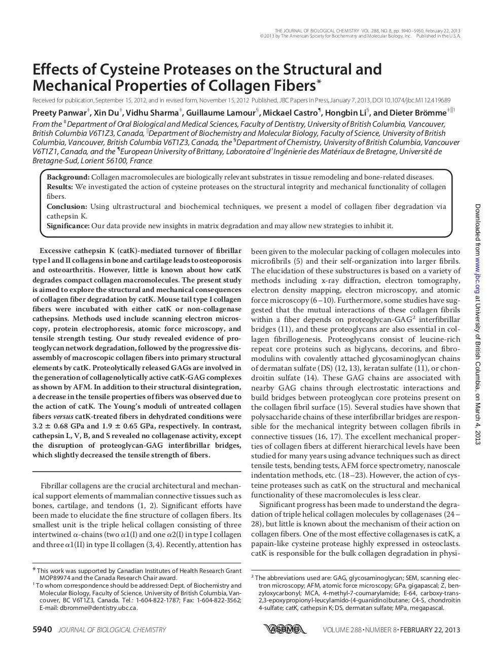
were constructed using reference proteins of different sizes.3
According to these standard curves, it was determined that the
apparent height of monomeric catK particles was 4.3 ⫾ 1.7 nm,
which is very similar to the observed catK size in the crystal
structure. Microscopic observation of catK-treated collagen
fibers exhibited the loss of surface interactions within 1 h and
thus the probable release of GAGs. This was further corroborated by the finding of increased levels of GAGs in catK-treated
collagen fibers (Fig. 3B). After 30 min of incubation with catK,
aggregates of potentially several catK molecules and GAGs
were observed with apparent heights of 5.5 ⫾ 1.1 nm (Fig. 4B).
This minor height difference is expected due to interaction
between catK and GAG strands forming larger size globular
complexes. Similar structures (Fig. 4B, arrow) were observed
when catK and C4-S were incubated alone (Fig. 4C). In contrast,
catK in the absence of C4-S provided single dot images as seen
in Fig. 4A (data not shown). After 4 h, fibrillar materials associated with globular aggregates appeared, and after 7 h, large
amount of fibrillar materials of different heights were visible
(Fig. 4, D and E). These fibrils likely correspond to tropocollagen aggregates and released fibrillar substructures accumulated
3
X. Du and D. Brömme, unpublished data.
5944 JOURNAL OF BIOLOGICAL CHEMISTRY
in the digest mixtures, which are subsequently degraded by
catK-GAG complexes. SDS-PAGE analysis of the solubilized
cleavage products revealed tropocollagen-sized ␣-chain-related bands of 100 –130 kDa. Thus, AFM images at 7 h represent released microfibrillar fragments but may also be interpreted as partially refibrilized soluble tropocollagen fragments
during sample preparation.
Degradation of Type I Collagen Fibers by catK in the Presence
of Additional GAGs and NaCl—Type I collagen fibers were
separately incubated with catK-C4-S and catK-DS mixtures (3
M catK ⫹ 30 g/ml) at 28 °C to determine the effects of externally applied GAGs on fibers degradation. Scanning microscopy analysis and in vitro digest data revealed no significant
differences in the degradation level of collagen fibers in the
presence or absence of external GAGs (Fig. 5, A–D). As the
degradation of collagen-associated proteoglycans by catK
releases sufficient amounts of GAGs, no externally added
GAGs are required for the degradation of native collagen fibers.
Adding 300 mM NaCl to the digest completely inhibited the
dissociation of the collagen fiber into smaller fibrils but did not
block the removal of GAGs. After 20 h of incubation, the collagen fibers stayed intact but showed an ⬃53% increase in fiber
diameter (Fig. 6E) and the release of GAGs (7.5 ⫾ 1.2 g) into
VOLUME 288 • NUMBER 8 • FEBRUARY 22, 2013
Downloaded from www.jbc.org at University of British Columbia, on March 4, 2013
FIGURE 4. AFM analysis of soluble fractions of type I collagen fiber degradation by catK. AFM images of supernatants of catK/type I collagen digests at 0-h
incubation ((A) only catK molecules of 4.3 ⫾ 1.7 nm height are visible; catK in buffer alone gives identical images (not shown)), 0.5-h incubation ((B) CatK-GAG
complexes of 5.5 ⫾ 1.1 nm height are present), and catK/collagen digests at 4-h incubation (D), and 7 h ((E) between 4 –7 h of increasing amounts of fibrillar
material of different heights appears). C, similar catK-GAG complexes as B (here, catK and C4-S were incubated in the absence of collagen at a catK:GAG ratio
similar to those present in the digestion supernatant after 30 min; 3 M catK:30 g/ml C4-S). The upper right insets are electronic magnifications of catK and
CatK-GAG complexes. The colored bars on the right in the upper and lower panels represent the height scale of A–E, respectively. Diameter measurements were
done by taking the average sample heights from different cross-sections. The black arrowheads point to catK molecules, and black arrows point to the catK-GAG
complexes.
Mechanism of Collagen Fiber Degradation by Cathepsin K
the digest mixture. This indicates that electrostatic interactions
are needed for the degradation of collagen but not for the degradation of collagen-associated proteoglycans. This is further
supported by the finding that in the presence of 300 mM NaCl
no ␣-band-sized fragments were observed in SDS-PAGE analysis (Fig. 7A, catK ⫹ NaCl lane). As NaCl at this concentration
does not have a significant effect on the kinetic parameters of
the hydrolysis of a fluorogenic peptide substrate or on the degradation of gelatin (denatured collagen) but prevents the complex formation with GAGs, it can be concluded that monomeric catK cleaves proteoglycans but does not cleave fibrillar
collagens as previously shown for the degradation of soluble
triple helical collagen (40).
Degradation of Insoluble Type I Collagen Fibers by Non-collagenolytic Cathepsins—SEM-based analysis of the collagen
fibers treated with different cathepsins revealed that cathepsins
B, S, and V only removed proteoglycan-GAG bridges between
fibrils (Fig. 6, A–D). SDS-PAGE data confirmed their lack of
collagenolytic activity. No collagen ␣-bands were observed. In
contrast to cathepsins B, S, and V, cathepsin L activity displayed
the removal of the proteoglycans and a partial liberation of
␣-band-sized fragments (Fig. 7A). SDS-PAGE analysis further
revealed the presence of very low amounts of lower molecular
FEBRUARY 22, 2013 • VOLUME 288 • NUMBER 8
weight sized ␣-chain-sized degradation products, indicating a
very weak collagenase activity. However, this should be carefully interpreted as this could be a consequence of the degradation of partially denatured tropocollagen molecules within the
fiber accessible for degradation by cathepsin L.
Effects of Cathepsins on Collagen Fiber Diameter and Weight—
There were minor changes in the diameters and weights of collagen fibers when incubated with activity buffer in the absence
of enzymes for up to 20 h. In contrast, incubation of fibers with
catK showed initial increases of fiber diameters from 70 –100%
after 2 h due to subsequent loss of fiber integrity. The complete
disappearance of fiber structure occurred after 14 h. The initial
increase in fiber diameter is likely attributed to the loss of interfibrillar GAG bridges, which may lead to a loosening of the fiber
structure. Less dramatic increases in fiber diameter were
observed by the action of non-collagenase cathepsins. Scanning
microscopy measurements showed ⬃62% enhancement in
diameter after 20 h of incubation with cathepsin L, ⬃45% with
cathepsin V or cathepsin B, ⬃59% with cathepsin S, and ⬃53%
with catK⫹NaCl (Fig. 7B). Weight determinations of digested
collagen fibers showed a 100% loss in mass after 14 h by catK,
demonstrating the complete digest of the fiber material.
Cathepsin L and catK ⫹ NaCl treated samples showed a ⬃35%
JOURNAL OF BIOLOGICAL CHEMISTRY
5945
Downloaded from www.jbc.org at University of British Columbia, on March 4, 2013
FIGURE 5. Degradation of type I collagen fibers by catK in the presence of C4-S. Shown are SEM micrographs of untreated collagen fiber (A), fibers
incubated with catK/C4-S for 4 h (B), and 10 h (C), showing the degradation of collagen fibers (45.5 ⫾ 6.5 m). The catK concentration is 3 M in presence of 30
g/ml C4-S at pH 5.5 and 28 °C. Bars represent 30 m. The structural degradation of collagen fiber shows a dramatic increase in diameter after 2 h, and their
dissociation into fibril bundles after 4 h is followed by their dissociation into subfibrillar structures. Between 7–14 h, the degradation of fibrils was clearly visible.
After 14 h, compact fibers disappeared as shown in catK degradation in the absence of external GAGs. In conclusion, the degradation of collagen fiber is similar
in both cases, in the absence or presence of endogenous C4-S. The released GAGs from collagen fibers by catK are sufficient for the formation of catK-GAG
complexes to degrade the compact collagen fiber and fibrils. D, SDS-PAGE analysis of collagen fiber degradation products (␣1- and ␣2-chains) after incubation
with 3 M catK in the presence of 30 g/ml C4-S at different time points up to 20 h. The time-dependent degradation pattern of ␣-bands is similar to that
observed in the absence of additional GAGs. Experiments performed with catK/DS mixtures showed identical results (not shown).
Mechanism of Collagen Fiber Degradation by Cathepsin K
FIGURE 6. Collagen fiber degradation by non-collagenolytic cathepsins.
SEM micrographs of collagen fibers after 20 h Incubation with cathepsin L (A),
cathepsin V (B), cathepsin B (C), cathepsin S (D), and catK⫹NaCl ((E) 3 M
enzyme concentrations) at pH 5.5 and room temperature. Left panel demonstrates the morphology of intact collagen fibers after 20-h cathepsin treatment; bars represent 30 m. Right panel shows the magnified surface view of
fibers after 20-h incubation with cathepsins L, V, B, S, and catK⫹NaCl. The
images clearly demonstrate the arrangement of fibrils after enzymatic action
and removal of collagen fiber-associated proteoglycans. Bars represent 2 m.
CatK in the presence of 300 mM NaCl and cathepsin L remove proteoglycans
and partially degrade the collagen fiber at its surface. Note the collagen fibers
incubated with activity buffer in the absence of cathepsins reveal no structural changes after 20 h.
5946 JOURNAL OF BIOLOGICAL CHEMISTRY
weight losses and cathepsins B-, S-, and V-treated samples
about a 20 –27% weight loss after 20 h of incubation (Fig. 7C).
Here, the weight loss is primarily related to the degradation of
proteoglycans and in the case of cathepsin L, a partial loss of
collagen is also likely as shown by the release of small amounts
of ␣-chain-related degradation products into the supernatants
of the degradation mixture.
Effects of Cysteine Proteases on Mechanical Properties of Collagen Fiber—The mechanical properties of collagen fibers vary
with different enzymatic treatments. The stress-strain curves
obtained from the tensile test of control fibers, catK, non-colVOLUME 288 • NUMBER 8 • FEBRUARY 22, 2013
Downloaded from www.jbc.org at University of British Columbia, on March 4, 2013
FIGURE 7. Cathepsin-mediated degradation of collagen fibers. A, comparative SDS-PAGE of collagen fiber degradation products after incubation with
activity buffer (C, control), catK, cathepsin L, cathepsin V, cathepsin B, cathepsin S, and catK⫹NaCl (enzyme concentration, 3 M) at pH 5.5 and 28 °C for 4 h,
respectively. Only catK is able to release significant amounts of tropocollagen
fragments from insoluble collagen fibers. B, diameter analysis of collagen
fibers after incubation with activity buffer (n ⫽ 14), catK, non-collagenase
cathepsins (cathepsins L, V, B, and S), and catK⫹NaCl (3 M enzyme concentration), respectively, at different time points (0 h, 2 h, and 20 h) (n ⫽ 8).
C, mass analysis of collagen fibers at different points (0 –20 h) of incubation
with activity buffer, catK, or non-collagenase cathepsins. As shown, incubation of collagen fibers with activity buffer in the absence of cathepsins does
not affect fiber diameters and their appearance, and no ␣-bands are observed
in SDS-PAGE. Note that K⫹NaCl represents the catK⫹NaCl.
Mechanism of Collagen Fiber Degradation by Cathepsin K
FIGURE 8. Mechanical strength measurements on collagen fibers after
cathepsin treatment. A, typical stress-strain curves of control collagen fibers
incubated with activity buffer in the absence of proteases for 20 h (n ⫽ 14).
Fibers were incubated with non-collagenase cathepsins (L, V, B, S, or
catK⫹NaCl) for 20 h (n ⫽ 8), and collagen fibers were incubated with catK (n ⫽
8) for 2 h. Stress-strain curves were obtained from the displacement of 5
mm/min in dry conditions. B, graph showing the Young’s moduli of control
collagen fibers and cathepsins K, L, V, B, S, and catK⫹ NaCl-treated fibers.
Young’s moduli were calculated on the linear part of the stress-strain curve.
The relative error in the tensile modulus of these fibers is due to the function
of different cathepsins and changes in fiber diameter. C, graph showing the
stress at break (f) and strain at break (‚) of control and cathepsin (K, L, V, B, S,
and catK⫹NaCl)-treated collagen fibers. Note that incubation time for catKtreated fibers is 2 h because after 2 h, fibers lost their structural integrity,
which interferes in diameter measurements. 2-h catK-treated fibers show
weak mechanical properties compared with other cathepsin-treated and
control fibers after 20 h.
lagenase cathepsins (L, V, B, and S) and catK⫹NaCl-treated
fibers are shown in Fig. 8A. The differences in the diameter of
fibers due to the specific activity of these proteases (Fig. 7B) are
the likely cause for the variation of the tensile strength properties of the fibers. The average Young’s moduli for control fibers
were 3.2 ⫾ 0.68 GPa, catK-treated fibers were 1.9 ⫾ 0.65 GPa
FEBRUARY 22, 2013 • VOLUME 288 • NUMBER 8
DISCUSSION
Model of Collagen Fiber Degradation by catK—Most studies
related to collagenases use soluble collagens as substrate,
although the biologically relevant substrates in tissue remodeling and diseases such as osteoporosis, arthritis, and fibroses are
insoluble collagen fibers or fibrils. Only recently, the fine structure of collagen fibrils has been better understood (41). The
morphology of collagen fibers is characterized by a regular
arrangement of fibrils tightly bundled together through GAGmediated proteoglycan interactions as shown in previous
reports (15, 39, 42, 43). We and others (24) have previously
demonstrated the unique collagenase activity of catK, which is
able to cleave at multiple sites within the triple helical region of
tropocollagen. But how is catK able to degrade collagen fibers
so efficiently? Our microscopic analysis and in situ proteolytic
digestion results of collagen fiber degradation by catK demonstrated that the dissociation of proteoglycans from the fibers
occurred prior to the collagen degradation and that their
removal will expose additional areas on the fiber surface for the
collagenolytic attack (44). However, the removal of proteoglycans alone does not appear to be sufficient to disintegrate a
macro fiber into smaller subfibrils, as cathepsin B and cathepsin
V are unable to do so and cathepsin L is only able to do so in a
limited manner (45, 46).
We have previously demonstrated that the collagenase activity of catK requires GAGs as cofactors (25). These GAGs are
likely provided by the degradation of collagen-associated proteoglycans to form collagenolytically active catK-GAG complexes (47). In this report, we demonstrated that catK releases
GAGs from collagen fibers, and this reaction correlates with the
release of collagen ␣-chains from the fiber. NaCl, which has
been previously shown to block soluble collagen degradation
and complex formation of catK with GAGs (40), completely
inhibits collagen fiber disassembly and degradation. Although
our data are indicative that NaCl prevents collagen degradation
by inhibiting the formation of collagenolytically active catKJOURNAL OF BIOLOGICAL CHEMISTRY
5947
Downloaded from www.jbc.org at University of British Columbia, on March 4, 2013
and for non-collagenase cathepsin (L, V, B, and S)-treated fibers
between 2.0 ⫾ 0.42 GPa to 2.7 ⫾ 0.76 GPa (Fig. 8B). However,
control fibers had significantly higher ultimate tensile strength
(2.13 ⫾ 0.71 N) than catK-treated fibers (0.95 ⫾ 0.55 N) and
non-collagenase cathepsin-treated fibers. The tensile results
are displayed in Table 1 and show that for control fibers, the
stress at the break is slightly higher (650 ⫾ 110 MPa) compared
with catK (241 ⫾ 121 MPa) and non-collagenase cathepsin
treated fibers (381 ⫾ 190 MPa to 556 ⫾ 86 MPa). Both, stress at
break and strain at failure were highest for control fibers and
lowest for catK-treated fibers (Fig. 8C). The maximum stretching was observed in control fibers (39 ⫾ 7%). On the other hand,
catK-treated collagen fibers had a strain at failure of 19 ⫾ 8%.
Approximately 27 ⫾ 4%, 25 ⫾ 3%, 29 ⫾ 6%, 31 ⫾ 7%, and 31 ⫾
4% stretching of collagen fibers were observed after incubation
with cathepsins L, V, B, S, and catK⫹NaCl, respectively. The
strain rate of collagen fibers decreased as a result of proteolytic
activity of non-collagenase cathepsins because these proteases
rupture the proteoglycan-GAG bridges between fibrils, which
affects the mechanical integrity between the fibrils of collagen
fibers (16, 17).
Mechanism of Collagen Fiber Degradation by Cathepsin K
TABLE 1
Mechanical properties of control and cathepsin-treated collagen fibers
Fiber treatment
Control fibers (n ⫽ 14)
CatK-treated fibers (n ⫽ 8)
CatL-treated fibers (n ⫽ 8)
CatV-treated fibers (n ⫽ 8)
CatB-treated fibers (n ⫽ 7)
CatS-treated fibers (n ⫽ 8)
CatK⫹NaCl-treated fibers (n ⫽ 8)
Diameter ⫾ S.D.a
m
60.03 ⫾ 15.56
81.31 ⫾ 22.71
76.02 ⫾ 17.22
70.09 ⫾ 18.25
65.95 ⫾ 7. 59
71.16 ⫾ 16.50
67.17 ⫾ 14.12
Strength ⫾ S.D.b
N
2.13 ⫾ 0.71
0.95 ⫾ 0.55
1.79 ⫾ 0.77
1.89 ⫾ 0.63
1.54 ⫾ 0.44
2.01 ⫾ 0.95
1.99 ⫾ 0.50
Young’s
modulus ⫾ S.D.
GPa
3.21 ⫾ 0.68
1.93 ⫾ 0.65
1.77 ⫾ 0.62
2.29 ⫾ 0.59
2.12 ⫾ 0.26
2.78 ⫾ 0.76
2.07 ⫾ 0.42
Stress at
break ⫾ S.D.
MPa
650 ⫾ 110
241 ⫾ 121
381 ⫾ 190
478 ⫾ 189
479 ⫾ 142
556 ⫾ 86
512 ⫾ 157
Strain at
failure ⫾ S.D.c
%
39 ⫾ 7
19 ⫾ 8
27 ⫾ 4
25 ⫾ 3
29 ⫾ 6
31 ⫾ 7
31 ⫾ 4
a
Average diameters of collagen fibers calculated from multiple spots on the same fiber in dry conditions.
Ultimate force in Newton (N).
c
Ultimate strain in percentage (%) representing the maximum stretching value till failure.
b
GAG complexes, it should be noted that we cannot rule out an
effect on the binding of catK to collagen by itself.
Our SEM data of catK-treated collagen fibers in the presence
of NaCl also provide direct evidence that in the absence of catKGAG complexes, monomeric catK still exerts its proteoglycan
degradation capability but lacks it collagen-degrading activity.
This suggests that catK-GAG complexes are only required for
the degradation of collagen fibrils as previously demonstrated
for soluble collagen fragments (25, 40). The stepwise degradation of collagen molecules from the surface of fibrils will allow
access to cryptic proteoglycans within the macro fiber core,
which will lead to the observed splitting of the fiber into smaller
and smaller subfibrils and simultaneously to an increased GAG
release by catK. As expected the progressive unfolding of the
fibers made them less stable and thus more accessible for further degradation. Scheme 1 summarizes the proposed mechanism of collagen fiber degradation by catK.
Mechanical Properties of Collagen Fibers Influenced by
Cathepsin Activity—Tendons are organized in a hierarchical
order from tropocollagen, microfibrils, and fibrils to fibers (4, 5,
41). Previous work has demonstrated the mechanical characterizations of different hierarchical structures of collagen fibers
(20, 21, 23, 48 –50). The comparison of undigested control collagen fibers with cathepsin-treated fibers in dehydrated conditions clearly indicated that the orientation, tensile strength, and
strain rate of collagen fibers changed due to the activity catK
5948 JOURNAL OF BIOLOGICAL CHEMISTRY
and non-collagenase cathepsins. As a consequence of cathepsin
activity, fiber diameter increased, and Young’s moduli and ultimately the tensile strength of collagen fibers decreased (Figs. 7B
and 8B). Our micromechanical results suggest that stress-strain
curves of control and cathepsin-treated collagen fibers exhibit
both linear and non-linear regions, which are in agreement with
previous studies (19, 21). We find that control collagen fibers
incubated in activity buffer up to 20 h and analyzed in dehydrated conditions had smaller diameters (⬃60 m) and displayed a higher Young’s modulus, ultimate strain, and strength.
However, an ⬃70% increase in diameter was observed after 2 h
of incubation time, leading ultimately to the complete degradation of collagen fibers by catK. The stress at the break, strain at
failure, Young’s modulus, and ultimate tensile strength of catKtreated collagen fibers was comparatively lower than control
and non-collagenase-treated fibers in dehydrated conditions
(Table 1).
The above results suggest that the control collagen fiber
appears to be more stable than catK and other non-collagenase
cathepsin-treated fibers. We believe that the load-bearing
mechanism between the collagen fibrils may disrupt due to
these enzymes. Fibrillar collagens in combination with proteoglycans form a network to provide mechanical integrity to
tissues (15–17). Proteoglycan/GAG interactions are mainly
responsible to hold fibrils together within a collagen fiber and
intermolecular cross-links in the telopeptide regions of triple
VOLUME 288 • NUMBER 8 • FEBRUARY 22, 2013
Downloaded from www.jbc.org at University of British Columbia, on March 4, 2013
SCHEME 1. Schematic presentation of stepwise collagen fiber degradation by catK. White and blue thin strands are proteoglycan-GAG chains, single red
dots are catK molecules, and aggregates of red dots are catK/GAG complexes.
Mechanism of Collagen Fiber Degradation by Cathepsin K
Acknowledgment—We are thankful to Antoine Kervoelen for kind
help in the mechanical experiments and for the technology support of
the Centre for High Throughput Phenogenomics.
REFERENCES
1. Prockop, D. J., and Kivirikko, K. I. (1995) Collagens: molecular biology,
diseases, and potentials for therapy. Annu. Rev. Biochem. 64, 403– 434
2. Kadler, K. E., Baldock, C., Bella, J., and Boot-Handford, R. P. (2007) Collagens at a glance. J. Cell Sci. 120, 1955–1958
3. Rich, A., and Crick, F. H. (1961) The molecular structure of collagen.
J. Mol. Biol. 3, 483–506
4. Okuyama, K., Takayanagi, M., Ashida, T., and Kakudo, M. (1977) A New
Structural Model for Collagen. Polymer J. 9, 341–343
5. Orgel, J. P., Irving, T. C., Miller, A., and Wess, T. J. (2006) Microfibrillar
structure of type I collagen in situ. Proc. Natl. Acad. Sci. U.S.A. 103,
9001–9005
6. Orgel, J. P., Miller, A., Irving, T. C., Fischetti, R. F., Hammersley, A. P., and
Wess, T. J. (2001) The in situ supermolecular structure of type I collagen.
Structure 9, 1061–1069
7. Zhang, G., Ezura, Y., Chervoneva, I., Robinson, P. S., Beason, D. P., Carine,
E. T., Soslowsky, L. J., Iozzo, R. V., and Birk, D. E. (2006) Decorin regulates
assembly of collagen fibrils and acquisition of biomechanical properties
during tendon development. J. Cell Biochem. 98, 1436 –1449
8. Raspanti, M., Ottani, V., and Ruggeri, A. (1989) Different architectures of
the collagen fibril: morphological aspects and functional implications. Int.
J. Biol. Macromol. 11, 367–371
9. Starborg, T., Lu, Y., Meadows, R. S., Kadler, K. E., and Holmes, D. F. (2008)
Electron microscopy in cell-matrix research. Methods 45, 53– 64
10. Baselt, D. R., Revel, J. P., and Baldeschwieler, J. D. (1993) Subfibrillar structure of type I collagen observed by atomic force microscopy. Biophysical
Journal 65, 2644 –2655
FEBRUARY 22, 2013 • VOLUME 288 • NUMBER 8
11. Scott, J. E. (1992) Supramolecular organization of extracellular matrix
glycosaminoglycans, in vitro and in the tissues. FASEB J. 6, 2639 –2645
12. Redaelli, A., Vesentini, S., Soncini, M., Vena, P., Mantero, S., and Montevecchi, F. M. (2003) Possible role of decorin glycosaminoglycans in fibril to
fibril force transfer in relative mature tendons-a computational study
from molecular to microstructural level. J. Biomech. 36, 1555–1569
13. Vesentini, S., Redaelli, A., and Montevecchi, F. M. (2005) Estimation of the
binding force of the collagen molecule-decorin core protein complex in
collagen fibril. J. Biomech. 38, 433– 443
14. Rees, S. G., Flannery, C. R., Little, C. B., Hughes, C. E., Caterson, B., and
Dent, C. M. (2000) Catabolism of aggrecan, decorin and biglycan in tendon. Biochem. J. 350, 181–188
15. Scott, J. E., Orford, C. R., and Hughes, E. W. (1981) Proteoglycan-collagen
arrangements in developing rat tail tendon. An electron microscopical
and biochemical investigation. Biochem. J. 195, 573–581
16. Puxkandl, R., Zizak, I., Paris, O., Keckes, J., Tesch, W., Bernstorff, S., Purslow, P., and Fratzl, P. (2002) Viscoelastic properties of collagen: synchrotron radiation investigations and structural model. Philos. Trans. R. Soc.
Lond. B. Biol. Sci. 357, 191–197
17. Liu, X., Yeh, M. L., Lewis, J. L., and Luo, Z. P. (2005) Direct measurement
of the rupture force of single pair of decorin interactions. Biochem. Biophys. Res. Commun. 338, 1342–1345
18. Gentleman, E., Lay, A. N., Dickerson, D. A., Nauman, E. A., Livesay, G. A.,
Dee, K. C. (2003) Mechanical characterization of collagen fibers and scaffolds for tissue engineering. Biomaterials 24, 3805–3813
19. Pins, G. D., Christiansen, D. L., Patel, R., Silver, F. H. (1997) Self-assembly
of collagen fibers. Influence of fibrillar alignment and decorin on mechanical properties. Biophys. J. 73, 2164 –2172
20. Eppell, S., Smith, B., Kahn, H., and Ballarini, R. (2006) Nano measurements
with micro-devices: mechanical properties of hydrated collagen fibrils. J.
Roy. Soc. Interface 3, 117–121
21. Silver, F. H., Freeman, J. W., and Seehra, G. P. (2003) Collagen self-assembly and the development of tendon mechanical properties. J. Biomech. 36,
1529 –1553
22. Yang, L., Fitié, C. F., van der Werf, K. O., Bennink, M. L., Dijkstra, P. J., and
Feijen, J. (2008) Mechanical properties of single electrospun collagen type
I fibers. Biomaterials 29, 955–962
23. van der Rijt, J. A., van der Werf, K. O., Bennink, M. L., Dijkstra, P. J., and
Feijen, J. (2006) Micromechanical Testing of Individual Collagen Fibrils.
Macromol. Biosci. 6, 697–702
24. Garnero, P. (1998) The Collagenolytic Activity of Cathepsin K Is Unique
among Mammalian Proteinases. J. Biol. Chem. 273, 32347–32352
25. Li, Z., Yasuda, Y., Li, W., Bogyo, M., Katz, N., Gordon, R. E., Fields, G. B.,
and Brömme, D. (2004) Regulation of collagenase activities of human
cathepsins by glycosaminoglycans. J. Biol. Chem. 279, 5470 –5479
26. Bertini, I., Fragai, M., Luchinat, C., Melikian, M., Toccafondi, M., Lauer,
J. L., and Fields, G. B. (2012) Structural Basis for Matrix Metalloproteinase
1-Catalyzed Collagenolysis. J. Am. Chem. Soc. 134, 2100 –2110
27. Sarkar, S. K., Marmer, B., Goldberg, G., and Neuman, K. C. (2012) SingleMolecule Tracking of Collagenase on Native Type I Collagen Fibrils Reveals Degradation Mechanism. Curr. Biol. 22, 1047–1056
28. Manka, S. W., Carafoli, F., Visse, R., Bihan, D., Raynal, N., Farndale, R. W.,
Murphy, G., Enghild, J. J., Hohenester, E., and Nagase, H. (2012) Structural
insights into triple-helical collagen cleavage by matrix metalloproteinase
1. Proc. Natl. Acad. Sci. U.S.A. 109, 12461–12466
29. Yamashita, D. S., and Dodds, R. A. (2000) Cathepsin K and the Design of
Inhibitors of Cathepsin K. Curr. Pharm. Des. 6, 1–24
30. Yasuda, Y., Kaleta, J., and Brömme, D. (2005) The role of cathepsins in
osteoporosis and arthritis: rationale for the design of new therapeutics.
Adv. Drug Deliv. Rev. 57, 973–93
31. Rajan, N., Habermehl, J., Coté, M. F., Doillon, C. J., and Mantovani, D.
(2006) Preparation of ready-to-use, storable and reconstituted type I collagen from rat tail tendon for tissue engineering applications. Nat. Protoc.
1, 2753–2758
32. Linnevers, C. J., McGrath, M. E., Armstrong, R., Mistry, F. R., Barnes,
M. G., Klaus, J. L., Palmer, J. T., Katz, B. A., and Brömme, D. (1997) Expression of human cathepsin K in Pichia pastoris and preliminary crystallographic studies of an inhibitor complex. Protein Sci. 6, 919 –921
JOURNAL OF BIOLOGICAL CHEMISTRY
5949
Downloaded from www.jbc.org at University of British Columbia, on March 4, 2013
helical collagen maintain the hierarchy of collagen molecules
within fibrils (5). From our analysis, it is clear that non-collagenase cathepsins (L, V, S, and B) are only capable of destabilizing
the fiber structure in a limited manner. Neither the proteoglycan degradation nor their previously described telopeptidase
activity (45, 46) allows for the disintegration of collagen fibers as
observed with catK. Therefore, the structures of collagen fibers
are still intact after incubation with non-collagenase cathepsins
up to 20 h but clearly displayed a decrease in the tensile strength
of fibers (19). However, catK possesses both proteoglycan and
triple helical collagen degradation abilities, which severely
interfere with the structural integrity as well as the mechanical
strength of collagen fibers.
In summary, we demonstrated the stepwise degradation of
collagen fibers by catK. SEM experiments revealed disintegration of large collagen fibers into smaller fibril bundles and
microfibrils and their final dissolution. This process is accompanied by the simultaneous degradation of collagen-associated
proteoglycans, which provide soluble GAGs to form collagenolytically active catK-GAG complexes. AFM studies suggested
the formation of defined catK-GAG complexes during the degradation process. Tensile strength studies showed the destabilizing effects of catK activity on the mechanical properties of
collagen fibers. However, we also investigated that destruction
of proteoglycan-collagen interactions by non-collagenase
cathepsins (catL, V, B, and S). These proteases are capable of
partially destabilizing collagen fibers by removing collagen-associated proteoglycans, which are reflected by the altered diameter of collagen fibers and decreased mechanical stability.
Mechanism of Collagen Fiber Degradation by Cathepsin K
5950 JOURNAL OF BIOLOGICAL CHEMISTRY
Hierarchical structures in fibrillar collagens. Micron 33, 587–596
42. Provenzano, P. P., and Vanderby, R., Jr. (2006) Collagen fibril morphology
and organization: implications for force transmission in ligament and tendon. Matrix Biol. 25, 71– 84
43. Paige, M. F., Rainey, J. K., and Goh, M. C. (2001) A study of fibrous long
spacing collagen ultrastructure and assembly by atomic force microscopy.
Micron 32, 341–353
44. Billinghurst, R. C., Wu, W., Ionescu, M., Reiner, A., Dahlberg, L., Chen, J.,
van Wart, H., and Poole, A. R. (2000) Comparison of the degradation of
type II collagen and proteoglycan in nasal and articular cartilages induced
by interleukin-1 and the selective inhibition of type II collagen cleavage by
collagenase. Arthritis Rheum. 43, 664 – 672
45. Burleigh, M. C., Barrett, A. J., and Lazarus, G. S. (1974) Cathepsin B1. A
lysosomal enzyme that degrades native collagen. Biochem. J. 137,
387–398
46. Kirschke, H., Kembhavi, A. A., Bohley, P., and Barrett, A. J. (1982) Action
of rat liver cathepsin L on collagen and other substrates. Biochem. J. 201,
367–372
47. Li, Z., Hou, W. S., and Brömme, D. (2000) Collagenolytic activity of cathepsin K is specifically modulated by cartilage-resident chondroitin sulfates. Biochemistry 39, 529 –536
48. Fratzl, P. (2008) Collagen: Structure and Mechanics, Springer, New York
49. Kato, Y. P., Christiansen, D. L., Hahn, R. A., Shieh, S. J., Goldstein, J. D., and
Silver, F. H. (1989) Mechanical properties of collagen fibres: a comparison
of reconstituted and rat tail tendon fibres. Biomaterials 10, 38 – 42
50. Gupta, H. S., Seto, J., Wagermaier, W., Zaslansky, P., Boesecke, P., and
Fratzl, P. (2006) Cooperative deformation of mineral and collagen in bone
at the nanoscale. Proc. Natl. Acad. Sci. 103, 17741–17746
VOLUME 288 • NUMBER 8 • FEBRUARY 22, 2013
Downloaded from www.jbc.org at University of British Columbia, on March 4, 2013
33. Brömme, D., Li, Z., Barnes, M., and Mehler, E. (1999) Human cathepsin V
functional expression, tissue distribution, electrostatic surface potential,
enzymatic characterization, and chromosomal localization. Biochemistry
38, 2377–2385
34. Brömme, D., Okamoto, K., Wang, B. B., and Biroc, S. (1996) Human cathepsin O2, a matrix protein-degrading cysteine protease expressed in
osteoclasts. Functional expression of human cathepsin O2 in Spodoptera
frugiperda and characterization of the enzyme. J. Biol. Chem. 271,
2126 –2132
35. Barrett, A. J., Kembhavi, A. A., Brown, M. A., Kirschke, H., Knight, C. G.,
Tamai, M., and Hanada, K. (1982) L-trans-Epoxysuccinyl-leucylamido(4guanidino)butane (E-64) and its analogues as inhibitors of cysteine proteinases including cathepsins B, H and L. Biochem. J. 201, 189 –198
36. Farndale, R. W., Sayers, C. A., and Barrett, A. J. (1982) A direct spectrophotometric microassay for sulfated glycosaminoglycans in cartilage cultures. Connect. Tissue Res. 9, 247–248
37. Tan, E. P., Ng, S. Y., and Lim, C. T. (2005) Tensile testing of a single
ultrafine polymeric fiber. Biomaterials 26, 1453–1456
38. Raspanti, M., Viola, M., Forlino, A., Tenni, R., Gruppi, C., and Tira, M. E.
(2008) Glycosaminoglycans show a specific periodic interaction with type
I collagen fibrils. J. Struct. Biol. 164, 134 –139
39. Orgel, J. P., Eid, A., Antipova, O., Bella, J., and Scott, J. E. (2009) Decorin
Core Protein (Decoron) Shape Complements Collagen Fibril Surface
Structure and Mediates Its Binding. PLoS One 4, e7028
40. Li, Z., Hou, W. S., Escalante-Torres, C. R., Gelb, B. D., and Bromme, D.
(2002) Collagenase activity of cathepsin K depends on complex formation
with chondroitin sulfate. J. Biol. Chem. 277, 28669 –28676
41. Ottani, V., Martini, D., Franchi, M., Ruggeri, A., and Raspanti, M. (2002)
Download 2013 panwar jbiolchem
2013_panwar_jbiolchem.pdf (PDF, 6.6 MB)
Download PDF
Share this file on social networks
Link to this page
Permanent link
Use the permanent link to the download page to share your document on Facebook, Twitter, LinkedIn, or directly with a contact by e-Mail, Messenger, Whatsapp, Line..
Short link
Use the short link to share your document on Twitter or by text message (SMS)
HTML Code
Copy the following HTML code to share your document on a Website or Blog
QR Code to this page

This file has been shared publicly by a user of PDF Archive.
Document ID: 0000094737.