Ambulatory Rheumatology, OA, Gout (PDF)
File information
Author: xenj
This PDF 1.5 document has been generated by Microsoft® Word 2010 Trial, and has been sent on pdf-archive.com on 11/06/2014 at 18:39, from IP address 121.54.x.x.
The current document download page has been viewed 850 times.
File size: 885.9 KB (8 pages).
Privacy: public file


File preview
Ambulatory Rheumatology, OA, Gout,
Tissue Rheumatism
Dr. N. Ferrer
Group 4 (SY 2013-2014)
INTRODUCTION
Five Major Categories of Rheumatic Diseases:
1. Soft tissue syndromes
- involves periarticular structures but primarily not
the joint itself
- muscles, ligaments, bones, nerves, tendons, bursa
Examples:
o Trigger finger (locking sound during
extension) – involves the tendon only. Assess
for inflammation. Examine contralateral area.
o Epicondylitis – pain in the elbow (involving
the medial/lateral epicondyle)
o Stiff neck – problem with muscles,
sternocleidomastoid
o Tenosynovitis – involves the tendon sheath
(e.g. De Quervain’s)
2. Degenerative joint diseases
Osteoarthritis
- not an inevitable consequence of aging, but
most likely to get it once we age. Not really
degenerative
- Major joint areas – large, weight bearing
joints (hips, knees, ankles. Response of certain
joint groups vary. Higher incidence in knee,
then hips. Ankles are spared most of the time.
3. Inflammatory rheumatic diseases
o Rheumatoid arthritis
- autoimmune, seronegative
o Spondylarthropathies
- less joint involvement but more of the spine
Inflammatory disorders may be :
o Infectious
Infection with Neisseria gonorrhoea or
Mycobacterium tuberculosis
o Crystal-induced
Gout
Pseudogout
o Immune-related
Rheumatoid arthritis
Systemic lupus erythematosus
o Reactive
Rheumatic fever
o
Reactive arthritis
Idiopathic
4. Acute monoarthritis
o Crystal deposition arthropathy
o Septic arthritis
5.
o
o
o
o
Oligoarthritis - < 4 joints involved
Polyarthritis - > 4 joints involved
Connectisve tissue diseases
Systemic lupus erythematosus
Dermatomyositis
Scleroderma
Systemic vasculitis
Worldwide Extent of Various Musculoskeletal Diseases
- 355 million people have arthritis
- Prevalence of RA and OA correlates with a
country’s level of industrial development
- By year 2025, degenerative bone and joint
disorders will be most common causes of physical
disability (up to 25% of all incapacitating
conditions)
- Socioeconomic impact of rheumatic conditions
increasing worldwide
Nonarticular Musculoskeletal Pain
o Tendinitis
o Bursitis
o Nerve Entrapment Syndromes
o Reflex Sympathetic Dystrophy
o Myofascial Pain
o Fibrositis (Fibromyalgia)
o Psychogenic
Nonarticular disorders
- Painful on active, but not passive (or assisted),
range of motion.
- Often demonstrate point or focal tenderness in
regions adjacent to articular structures, and have
physical findings remote from the joint capsule.
- Seldom demonstrate swelling, crepitus, instability,
or deformity of the joint itself.
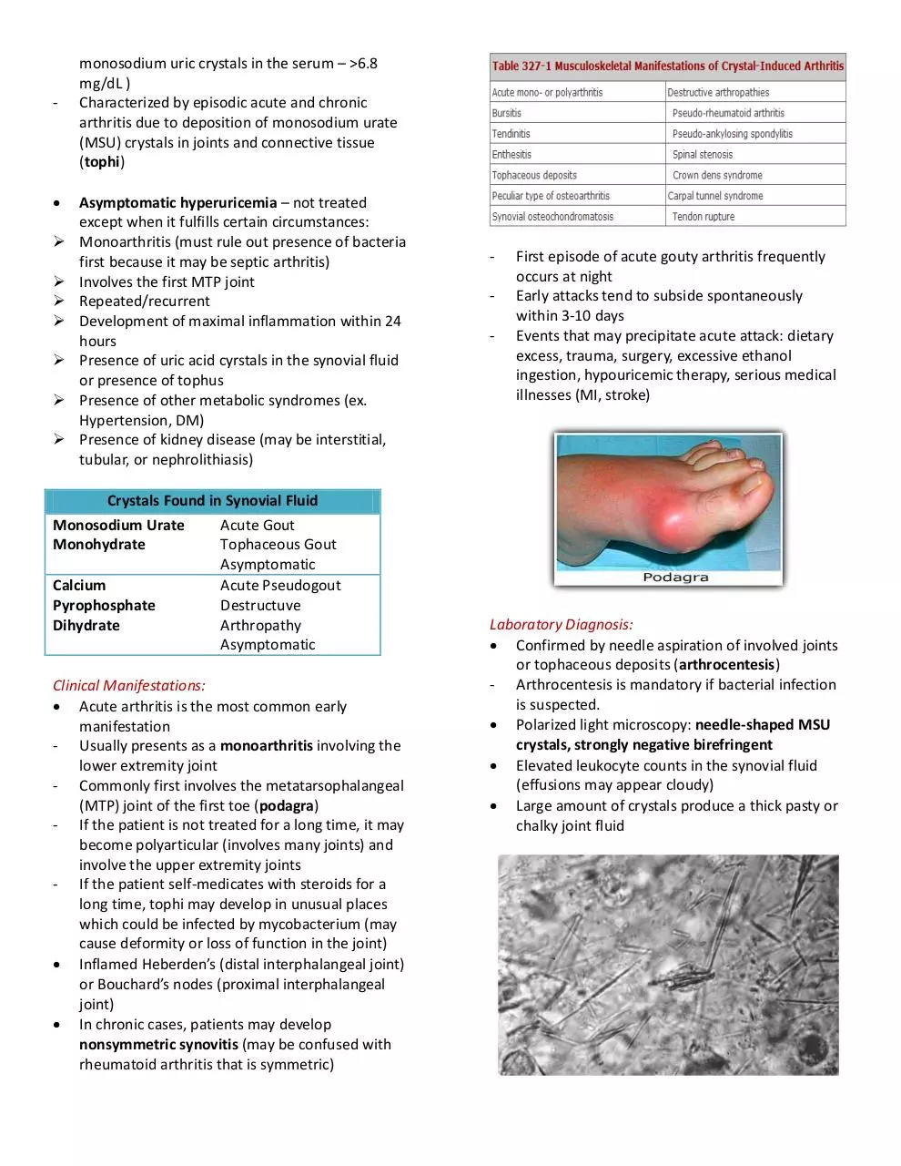
GOUT
- A group of diseases which presents as
hyperuricemia, acute arthritis, renal problems
(nephrolithiasis)
- Most often affects middle-aged to elderly men
and postmenopausal women
- Primary problem: hyperuricemia (saturation of
-
monosodium uric crystals in the serum – >6.8
mg/dL )
Characterized by episodic acute and chronic
arthritis due to deposition of monosodium urate
(MSU) crystals in joints and connective tissue
(tophi)
Asymptomatic hyperuricemia – not treated
except when it fulfills certain circumstances:
Monoarthritis (must rule out presence of bacteria
first because it may be septic arthritis)
Involves the first MTP joint
Repeated/recurrent
Development of maximal inflammation within 24
hours
Presence of uric acid cyrstals in the synovial fluid
or presence of tophus
Presence of other metabolic syndromes (ex.
Hypertension, DM)
Presence of kidney disease (may be interstitial,
tubular, or nephrolithiasis)
Crystals Found in Synovial Fluid
Monosodium Urate
Acute Gout
Monohydrate
Tophaceous Gout
Asymptomatic
Calcium
Acute Pseudogout
Pyrophosphate
Destructuve
Dihydrate
Arthropathy
Asymptomatic
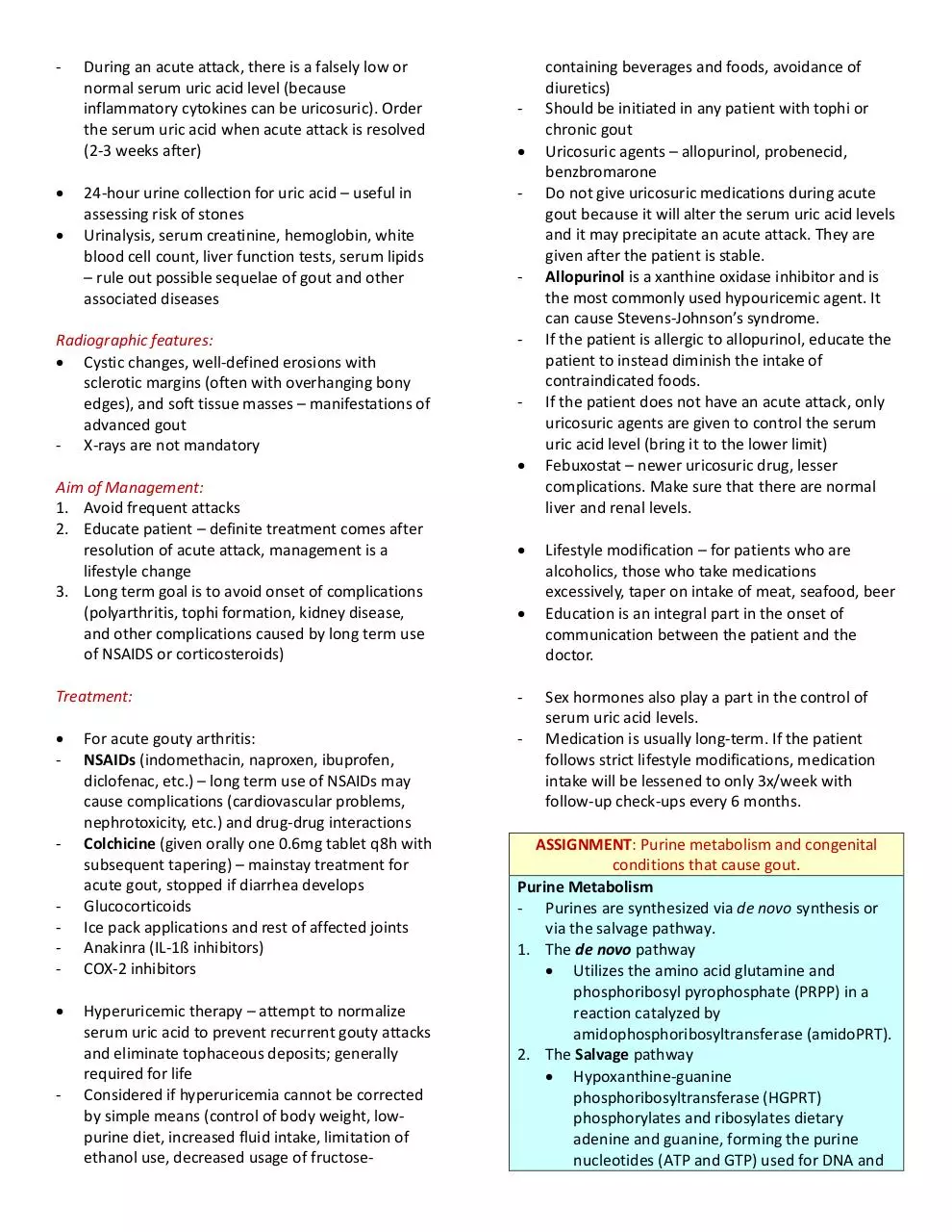
Clinical Manifestations:
Acute arthritis is the most common early
manifestation
- Usually presents as a monoarthritis involving the
lower extremity joint
- Commonly first involves the metatarsophalangeal
(MTP) joint of the first toe (podagra)
- If the patient is not treated for a long time, it may
become polyarticular (involves many joints) and
involve the upper extremity joints
- If the patient self-medicates with steroids for a
long time, tophi may develop in unusual places
which could be infected by mycobacterium (may
cause deformity or loss of function in the joint)
Inflamed Heberden’s (distal interphalangeal joint)
or Bouchard’s nodes (proximal interphalangeal
joint)
In chronic cases, patients may develop
nonsymmetric synovitis (may be confused with
rheumatoid arthritis that is symmetric)
-
First episode of acute gouty arthritis frequently
occurs at night
Early attacks tend to subside spontaneously
within 3-10 days
Events that may precipitate acute attack: dietary
excess, trauma, surgery, excessive ethanol
ingestion, hypouricemic therapy, serious medical
illnesses (MI, stroke)
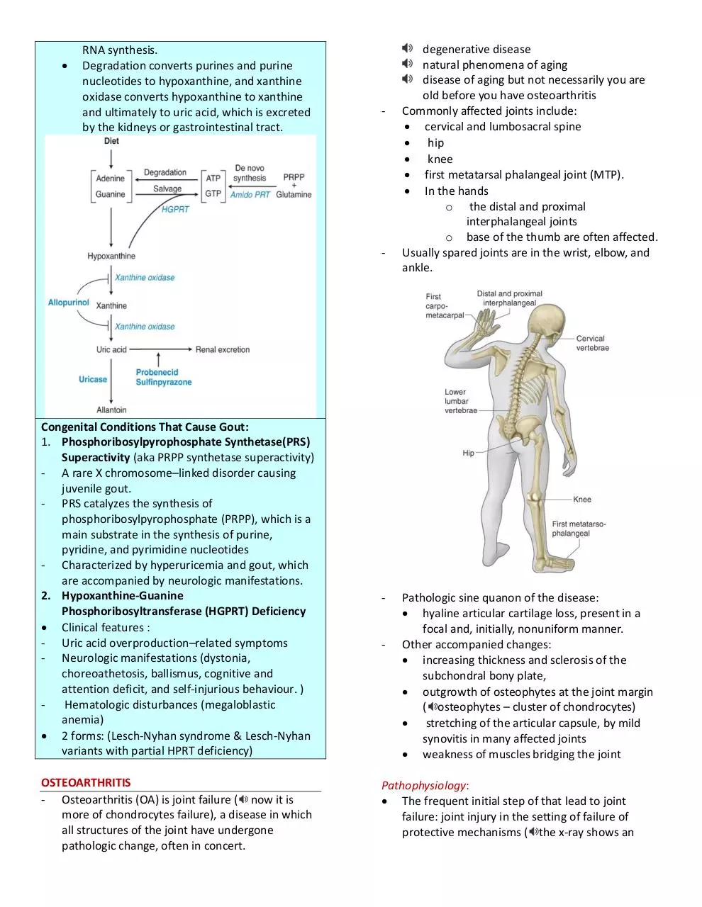
Laboratory Diagnosis:
Confirmed by needle aspiration of involved joints
or tophaceous deposits (arthrocentesis)
- Arthrocentesis is mandatory if bacterial infection
is suspected.
Polarized light microscopy: needle-shaped MSU
crystals, strongly negative birefringent
Elevated leukocyte counts in the synovial fluid
(effusions may appear cloudy)
Large amount of crystals produce a thick pasty or
chalky joint fluid
-
During an acute attack, there is a falsely low or
normal serum uric acid level (because
inflammatory cytokines can be uricosuric). Order
the serum uric acid when acute attack is resolved
(2-3 weeks after)
24-hour urine collection for uric acid – useful in
assessing risk of stones
Urinalysis, serum creatinine, hemoglobin, white
blood cell count, liver function tests, serum lipids
– rule out possible sequelae of gout and other
associated diseases
Radiographic features:
Cystic changes, well-defined erosions with
sclerotic margins (often with overhanging bony
edges), and soft tissue masses – manifestations of
advanced gout
- X-rays are not mandatory
Aim of Management:
1. Avoid frequent attacks
2. Educate patient – definite treatment comes after
resolution of acute attack, management is a
lifestyle change
3. Long term goal is to avoid onset of complications
(polyarthritis, tophi formation, kidney disease,
and other complications caused by long term use
of NSAIDS or corticosteroids)
-
-
-
Treatment:
-
-
-
-
-
For acute gouty arthritis:
NSAIDs (indomethacin, naproxen, ibuprofen,
diclofenac, etc.) – long term use of NSAIDs may
cause complications (cardiovascular problems,
nephrotoxicity, etc.) and drug-drug interactions
Colchicine (given orally one 0.6mg tablet q8h with
subsequent tapering) – mainstay treatment for
acute gout, stopped if diarrhea develops
Glucocorticoids
Ice pack applications and rest of affected joints
Anakinra (IL-1ß inhibitors)
COX-2 inhibitors
Hyperuricemic therapy – attempt to normalize
serum uric acid to prevent recurrent gouty attacks
and eliminate tophaceous deposits; generally
required for life
Considered if hyperuricemia cannot be corrected
by simple means (control of body weight, lowpurine diet, increased fluid intake, limitation of
ethanol use, decreased usage of fructose-
containing beverages and foods, avoidance of
diuretics)
Should be initiated in any patient with tophi or
chronic gout
Uricosuric agents – allopurinol, probenecid,
benzbromarone
Do not give uricosuric medications during acute
gout because it will alter the serum uric acid levels
and it may precipitate an acute attack. They are
given after the patient is stable.
Allopurinol is a xanthine oxidase inhibitor and is
the most commonly used hypouricemic agent. It
can cause Stevens-Johnson’s syndrome.
If the patient is allergic to allopurinol, educate the
patient to instead diminish the intake of
contraindicated foods.
If the patient does not have an acute attack, only
uricosuric agents are given to control the serum
uric acid level (bring it to the lower limit)
Febuxostat – newer uricosuric drug, lesser
complications. Make sure that there are normal
liver and renal levels.
Lifestyle modification – for patients who are
alcoholics, those who take medications
excessively, taper on intake of meat, seafood, beer
Education is an integral part in the onset of
communication between the patient and the
doctor.
Sex hormones also play a part in the control of
serum uric acid levels.
Medication is usually long-term. If the patient
follows strict lifestyle modifications, medication
intake will be lessened to only 3x/week with
follow-up check-ups every 6 months.
ASSIGNMENT: Purine metabolism and congenital
conditions that cause gout.
Purine Metabolism
- Purines are synthesized via de novo synthesis or
via the salvage pathway.
1. The de novo pathway
Utilizes the amino acid glutamine and
phosphoribosyl pyrophosphate (PRPP) in a
reaction catalyzed by
amidophosphoribosyltransferase (amidoPRT).
2. The Salvage pathway
Hypoxanthine-guanine
phosphoribosyltransferase (HGPRT)
phosphorylates and ribosylates dietary
adenine and guanine, forming the purine
nucleotides (ATP and GTP) used for DNA and
RNA synthesis.
Degradation converts purines and purine
nucleotides to hypoxanthine, and xanthine
oxidase converts hypoxanthine to xanthine
and ultimately to uric acid, which is excreted
by the kidneys or gastrointestinal tract.
-
-
Congenital Conditions That Cause Gout:
1. Phosphoribosylpyrophosphate Synthetase(PRS)
Superactivity (aka PRPP synthetase superactivity)
- A rare X chromosome–linked disorder causing
juvenile gout.
- PRS catalyzes the synthesis of
phosphoribosylpyrophosphate (PRPP), which is a
main substrate in the synthesis of purine,
pyridine, and pyrimidine nucleotides
- Characterized by hyperuricemia and gout, which
are accompanied by neurologic manifestations.
2. Hypoxanthine-Guanine
Phosphoribosyltransferase (HGPRT) Deficiency
Clinical features :
- Uric acid overproduction–related symptoms
- Neurologic manifestations (dystonia,
choreoathetosis, ballismus, cognitive and
attention deficit, and self-injurious behaviour. )
Hematologic disturbances (megaloblastic
anemia)
2 forms: (Lesch-Nyhan syndrome & Lesch-Nyhan
variants with partial HPRT deficiency)
OSTEOARTHRITIS
- Osteoarthritis (OA) is joint failure ( now it is
more of chondrocytes failure), a disease in which
all structures of the joint have undergone
pathologic change, often in concert.
-
-
degenerative disease
natural phenomena of aging
disease of aging but not necessarily you are
old before you have osteoarthritis
Commonly affected joints include:
cervical and lumbosacral spine
hip
knee
first metatarsal phalangeal joint (MTP).
In the hands
o the distal and proximal
interphalangeal joints
o base of the thumb are often affected.
Usually spared joints are in the wrist, elbow, and
ankle.
Pathologic sine quanon of the disease:
hyaline articular cartilage loss, present in a
focal and, initially, nonuniform manner.
Other accompanied changes:
increasing thickness and sclerosis of the
subchondral bony plate,
outgrowth of osteophytes at the joint margin
( osteophytes – cluster of chondrocytes)
stretching of the articular capsule, by mild
synovitis in many affected joints
weakness of muscles bridging the joint
Pathophysiology:
The frequent initial step of that lead to joint
failure: joint injury in the setting of failure of
protective mechanisms ( the x-ray shows an
osteophytes that are trying to compensate for the
cartilage thining or erosion.)
Joint Protectors and their significance in OA
Joint protectors
Function
Joint capsule and
ligaments
Synovial fluid
Ligaments, skin
and tendons
overlying the
joints
It limits the excursion of joints,
thereby fixing the range of
motion
Reduces friction between the
two articulating cartilage
surfaces thereby serving as
major protector against frictioninduced cartilage wear.
Contain mechanoreceptor
sensory afferent nerves;
anticipate joint loading
Muscles and
tendons
-
Key joint protector; provide the
appropriate power and
acceleration for the limb to
accomplish its task.
Failure of joint protectors would lead to increases
the risk in joint injury and OA.
Examples:
Charcot arthropathy – a severe and rapidly
progressive OA, develops when minor joint
injury occurs in the presence of posterior
column peripheral neuropathy.
Rupture of ligaments, a well-known cause of
the early development of OA.
Please refer to Harrison’s for bigger pic.
Normal cartilage matrix synthesis and catabolism:
1. In normal cartilage, type 2 collagen is woven tightly,
constraining the aggrecan molecules in the interstices
between collagen strands, forcing these highly
negatively charged molecules into close proximity
with one another.
2. The aggrecan molecule, through electrostatic
repulsion of its negative charges, gives cartilage its
compressive stiffness
3. Chondrocytes, the cells within this avascular tissue,
synthesize all elements of the matrix. In addition, they
produce enzymes that break down the matrix and
cytokines and growth factors, which in turn provide
autocrine/paracrine feedback that modulates
synthesis of matrix molecules
4. Cartilage matrix synthesis and catabolism are in a
dynamic equilibrium influenced by the cytokine and
growth factor environment.
Disruption in the equilibrium in OA:
1. Mechanical and osmotic stress will alter gene
expression on chondrocytes
2. Chondrocytes will produce more inflammatory
cytokines and matrix degrading enzymes such as:
collagenase (MMP-13) for Type 2 collagen
degradation, aggrecanases (ADAMTS-4 and ADAMTS5) for aggrecan. In which there is imbalance between
the production of the degrading enzymes and the
production of the elements of matrix as well at the
enzyme that promotes chondrocytes anabolism.
3. These degrading enzymes will eventually spread
throughout the matrix especially in superficial layer.
4. In synovium, there are also cytokines and growth
factors that are produce/synthesize. The following are
the enzymes produce that help chondrocytes
anabolism: BMP-2 (bone morphogenetic protein-2),
tissue inhibitor of metalloproteinase (TIMP), insulinlike growth factor type 1 and transforming growth
factor β. On the other hand, these are the
chondrocyte degrading enzyme: IL-1 (inteleukin-1)
and TNFα (tumor necrosis factor- α)
5. OA cartilage is characterized by gradual depletion of
aggrecan, an unfurling of the tightly woven collagen
matrix, and loss of type 2 collagen. With these
changes comes increasing vulnerability of cartilage,
which loses its compressive stiffness.
Risk Factors:
Joint vulnerability and joint loading are the two
major factors contributing to the development of
OA.
1. Systemic Risk Factors - Age
Most potent risk factor for OA
Aged cartilage is less responsive to these stimuli.
Aging increases the likelihood of failure of major
joint protectors.
Sensory nerve input slows with age, retarding the
feedback loop of mechanoreceptors to muscles
and tendons related to their tension and position.
Ligaments stretch with age, making them less able
to absorb impulses.
Older women are at high risk of OA in all joints.
Hormone loss with menopause may contribute to
this risk.
2. Heritability and Genetics
Highly heritable disease, but heritability varies by
joint.
The gene implicated is FRZB, a mutation may put
a woman at high risk of hip OA.
3. Global Considerations
Hip OA is rare in China and in immigrants from
China to the United States while knee OA is at
least as commonin Chinese than in Caucasians
from the United States, and knee OA represents a
major cause of disability in China.
Anatomic differences between Chinese and
Caucasian hips may account for much of the
difference in prevalence.
4. Risk Factors in the Joint Environment
Three uncommon developmental abnormalities
occurring in utero or childhood:
o Congenital Dysplasia, Legg-Perthes disease,
and Slipped Femoral Capital Epiphysis.
Girls are predominantly affected by acetabular
dysplasia, a mild form of congenital dislocation,
whereas the other abnormalities more often
affect boys.
Major injuries to a joint also can produce
anatomic abnormalities that leave the joint
susceptible to OA such as a fracture or avascular
necrosis.
Tears of ligaments that protect the joints, such as
the anterior cruciate ligament in the knee and the
labrum in the hip, can increase joint susceptibility
and lead to premature OA.
Malalignment across the joint. Varus (bowlegged)
knees with OA are at exceedingly high risk of
cartilage loss in the medial or inner compartment
of the knee, whereas valgus (knock-kneed)
malalignment predisposes to rapid cartilage loss
in the lateral compartment.
5. Loading Factors
A. Obesity
Three to six times body weight is exerted across
the knee during single leg stance.
Obesity is a well-recognized and potent risk factor
for the development of knee OA and, less so, for
hip OA.
It is a stronger risk factor for disease in women
than in men, and in women, the relationship of
weight to the risk of disease is linear.
B. Repeated Use of Joint
Occupational use and leisure time physical
activities.
Workers performing repetitive tasks as part of
their occupations for many years are at high risk
of developing OA in the joints they use
repeatedly.
While exercise is a major element of the
treatment of OA, certain types of exercise may
paradoxically increase the risk of disease.
Panarticular involvement of disease.
Cartilage initially shows surface fibrillation and
irregularity.
Focal erosions develop there, and these
eventually extend down to the subjacent bone.
Cartilage erosion down to bone expands to
involve a larger proportion of the joint surface,
even though OA remains a focal disease with
nonuniform loss of cartilage.
Changes:
Thinning of the Cartilage
Presence of Osteophyte
Subchondral Sclerosis
Asymmetrical Erosions
Sources of Pain:
Cartilage is aneural so cartilage loss in a joint is
not accompanied by pain.
Pain in OA likely arises from structures outside
the cartilage including the synovium, ligaments,
joint capsule, muscles, and subchondral bone.
Most of these are not visualized by the x-ray, and
the severity of x-ray changes in OA correlates
poorly with pain severity.
Clinical Features:
Pain is activity-related; it comes on either during
or just after joint use and then gradually resolves.
Early in disease, pain is episodic, triggered often
by a day or two of overactive use of a diseased
joint. Then the pain becomes continuous and
even begins to be bothersome at night.
Stiffness of the affected joint may be prominent,
but morning stiffness is usually brief.
In the knee, pain with activities requiring knee
flexion such as stair climbing.
A physical examination should focus on whether
tenderness is over the joint line (at the junction of
the two bones around which the joint is
articulating) or is outside of it.
No blood tests are routinely indicated for workup
of patients with OA unless less symptoms and
signs suggest inflammatory arthritis.
Examination of the synovial fluid is often more
helpful diagnostically than an x-ray.
o If the synovial fluid white count is >1000
per L, inflammatory arthritis or gout or
pseudogout are likely, the latter two
being also identified by the presence of
crystals.
X-rays are indicated to evaluate chronic hand pain
and hip pain thought to be due to OA, as the
diagnosis is often unclear without confirming
radiographs.
o For knee pain, x-rays should be obtained
if symptoms or signs are not typical of OA
or if knee pain persists after effective
treatment.
Radiographic findings correlate poorly with the
presence and severity of pain. Radiographs may
be normal in early disease as they are insensitive
to cartilage loss and other early findings.
Differential Diagnosis:
OA is the most common cause of chronic knee
pain in persons over age 45.
Inflammatory arthritis - prominent morning
stiffness and many other joints are affected.
Bursitis - occurs commonly around knees and
hips.
Anserine bursitis (medial and distal to the knee) is an extremely common cause of chronic knee
pain that may respond to a glucocorticoid
injection.
Trochanteric Bursitis –pain is isolated to an area
lateral to the hip joint.
Treatment:
• Treatment goal—alleviate pain and minimize loss of
physical function.
• Nonpharmacotherapy strategies aimed at altering
loading across the painful joint— includes patient
education, weight reduction, appropriate use of cane
and other supports, isometric exercises to strengthen
muscles around affected joints, bracing/orthotics to
correct malalignment.
• Topical capsaicin cream may help relieve hand or
knee pain.
• Acetaminophen, salicylates, NSAIDs, COX-2
inhibitors—must weigh individual risks and benefits.
• Tramadol—may be considered in patients whose
symptoms are inadequately controlled with NSAIDs;
as it is a synthetic opioid agonist, habituation is a
potential concern.
• Intraarticular glucocorticoids—may provide
symptomatic relief but typically short-lived.
• Intraarticular hyaluronin—can be given for
symptomatic knee and hip OA but it is controversial
whether they have efficacy beyond placebo.
• Glucosamine and chondroitin—although widely
sold, is not FDA approved for use in OA. Proof of
efficacy has not been established.
• Systemic glucocorticoids have no place in the
treatment of OA.
• Arthroscopic debridement and lavage—can be
helpful in the subgroup of patients with knee OA in
whom disruption of the meniscus causes mechanical
symptoms such as locking or buckling. In patients who
do not have mechanical symptoms, this modality
appears to be of no greater benefit than placebo.
• Joint replacement surgery may be considered in
patients with advanced OA who have intractable pain
and loss of function in whom aggressive medical
management has failed.
ASSIGNMENT: What are the biochemical changes
seen in the joints in aging and in osteoarthritis?
Age-Relating Mechanisms Triggering OA:
Mechanism
Oxidative stress
Apoptosis/autophagy
Matrix protein modification
(e.g., glycosylation)
Nonenzymatic collagen
cross-linking
Sarcopenia
Loss of proprioception
Joint laxity
Synovitis
Increased osteon density
Reduced circulating
progenitor cells (e.g., MSC)
Consequence
Cell senescence
Reduced regeneration
capacity
Reduced elasticity, fluid
content, stability
Impaired crack-bridging
potency
Reduced joint stability
Microtrauma, Charcotlike arthropathy
Microtrauma
Inflammatory cytokine
production
Bone stiffness
Impaired regeneration
capacity
The mechanisms leading to OA are complex and not
yet clear, but chondrocytes are at the center of the
process, which can be divided into several phases:
1. Chondrocyte injury, which is related to aging and
genetic and biochemical factors;
2. Early OA, in which chondrocytes proliferate
(cloning) and secrete inflammatory mediators,
collagens, proteoglycans, and proteases, which act
together to remodel the cartilaginous matrix and
initiate secondary inflammatory changes in the
synovium and subchondral bone; and
3. Late OA, in which repetitive injury and chronic
inflammation lead to chondrocyte drop out, marked
loss of cartilage, and extensive subchondral bone
changes.
- GOOD LUCK Notes by: Alcantara E, Carvajal K, Dizor J, Iwag M,
Lumasag J, Prado M, Sameon N
References: audio, JAX notes, Harrison’s 18th Ed.
Download Ambulatory Rheumatology, OA, Gout
Ambulatory Rheumatology, OA, Gout.pdf (PDF, 885.9 KB)
Download PDF
Share this file on social networks
Link to this page
Permanent link
Use the permanent link to the download page to share your document on Facebook, Twitter, LinkedIn, or directly with a contact by e-Mail, Messenger, Whatsapp, Line..
Short link
Use the short link to share your document on Twitter or by text message (SMS)
HTML Code
Copy the following HTML code to share your document on a Website or Blog
QR Code to this page

This file has been shared publicly by a user of PDF Archive.
Document ID: 0000168427.