Die vordere und seitliche Halsregion ohne Lücken (PDF)
File information
This PDF 1.4 document has been generated by Draw / LibreOffice 4.2, and has been sent on pdf-archive.com on 03/07/2014 at 00:07, from IP address 95.223.x.x.
The current document download page has been viewed 3765 times.
File size: 3.36 MB (55 pages).
Privacy: public file


File preview
Die vordere und
seitliche Halsregion
Prof. Dr. Carsten Theiss
!
Institut für Anatomie und Molekulare Embryologie
Leitung:
Abteilung für Cytologie
Hals
Grenzen:
cranialMandibula, Spitze des Proc.
mastoideus, Protuberantia
occ.ext.
!
caudalJugulum sterni, Clavicula,
Acromion, Proc. spinosus des
7. HW
Aus Prometheus
li. 1.Schicht der
Nackenmuskulatur:
!
M. trapezius
!!
re. 2. Schicht der
Nackenmuskulatur:
!
M.splenius
cervicis et
capitis
Aus Rohen/Yokochi
M. longissimus capitis
3. Schicht der
Nackenmuskulatur
M. semispinalis
capitis
4. Schicht der
Nackenmuskulatur
!
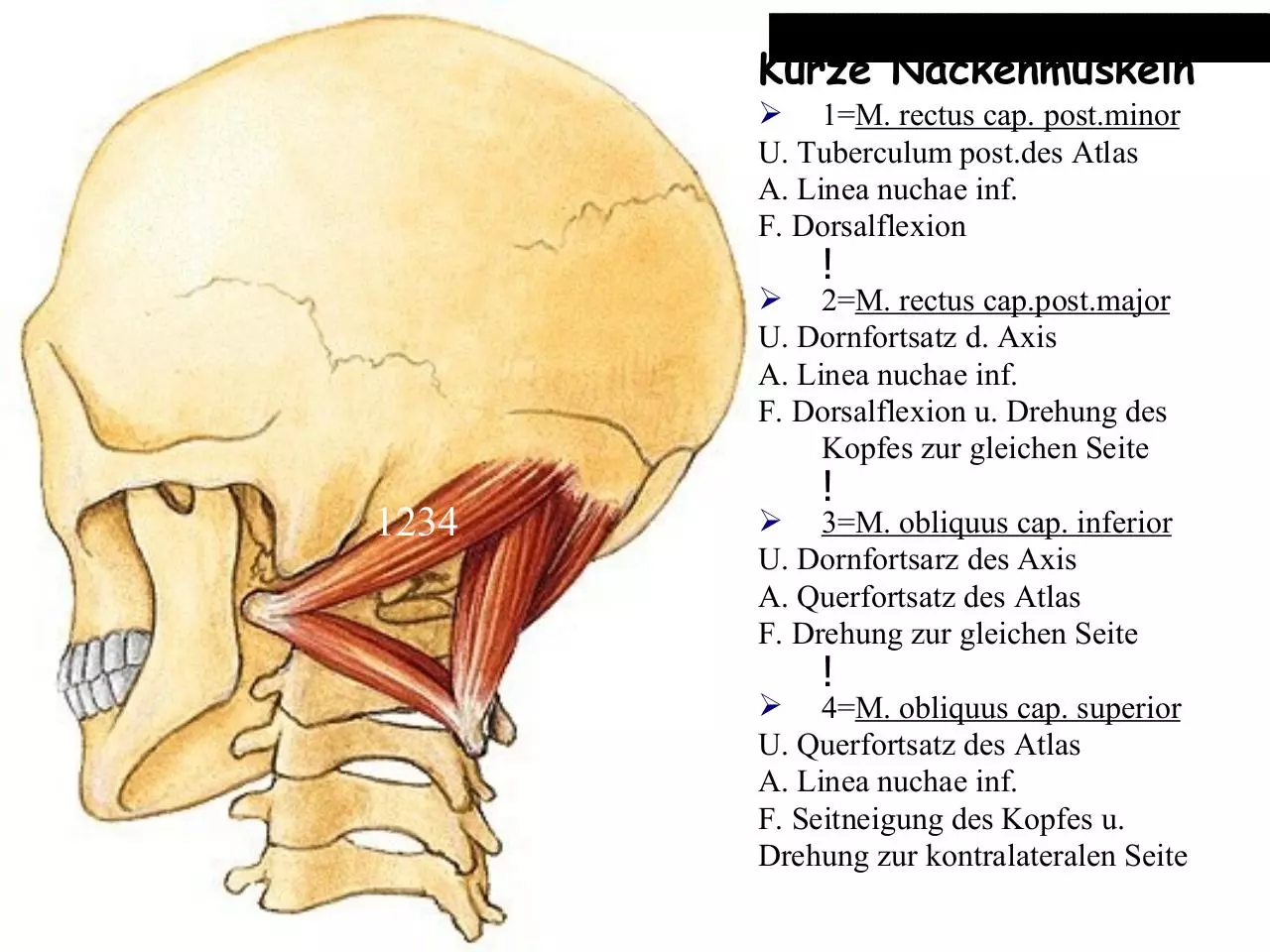
kurze Nackenmuskeln
M. multifidus
M. semispinalis
cervicis.
Aus Sobotta
Kurze Nackenmuskeln
➢ 1=M. rectus cap. post.minor
U. Tuberculum post.des Atlas
A. Linea nuchae inf.
F. Dorsalflexion
!
➢ 2=M. rectus cap.post.major
U. Dornfortsatz d. Axis
A. Linea nuchae inf.
F. Dorsalflexion u. Drehung des
Kopfes zur gleichen Seite
1234
!
➢ 3=M. obliquus cap. inferior
U. Dornfortsarz des Axis
A. Querfortsatz des Atlas
F. Drehung zur gleichen Seite
!
➢ 4=M. obliquus cap. superior
U. Querfortsatz des Atlas
A. Linea nuchae inf.
F. Seitneigung des Kopfes u.
Drehung zur kontralateralen Seite
Platysma
ist ein Hautmuskel
(epifaszial)
bewegt kein Gelenk
spannt die Haut des
Halses
Aus Lanz/Wachsmuth
Halsfaszien
Prometheus
Lamina superficialis
• oberflächliches Blatt
!
• Teil der allgemeinen Körperfaszie
!
• grenzt das Unterhautfettgewebe gegen die
Muskulatur des Bewegungsapparates ab
!
• umschließt (beide Muskeln liegen in der Faszie)
• M. sternocleidomastoideus
• M. trapezius
Oberflächliche Halsmuskeln:
(Platysma)
M. sternocleidomastoideus
M. trapezius
Prometheus
Download Die vordere und seitliche Halsregion ohne Lücken
Die vordere und seitliche Halsregion ohne Lücken.pdf (PDF, 3.36 MB)
Download PDF
Share this file on social networks
Link to this page
Permanent link
Use the permanent link to the download page to share your document on Facebook, Twitter, LinkedIn, or directly with a contact by e-Mail, Messenger, Whatsapp, Line..
Short link
Use the short link to share your document on Twitter or by text message (SMS)
HTML Code
Copy the following HTML code to share your document on a Website or Blog
QR Code to this page

This file has been shared publicly by a user of PDF Archive.
Document ID: 0000171998.