G6PD is a typical cytoplasmic (PDF)
File information
This PDF 1.5 document has been generated by convertonlinefree.com, and has been sent on pdf-archive.com on 25/02/2015 at 23:30, from IP address 41.204.x.x.
The current document download page has been viewed 781 times.
File size: 449.94 KB (10 pages).
Privacy: public file


File preview
GLUCOSE 6-PHOSPHATE DEHYDROGENASE DEFICIENCY
RELATIONSHIP WITH MALARIA
REVIEWED BY
YANKEY JUSTICE KWASI
24/11/2014
AND IT`S
GLUCOSE 6-PHOSPHATE DEHYDROGENASE (G6PD) DEFICIENCY
AND IT`S RELATIONSHIP WITH MALARIA
Glucose 6-phosphate dehydrogenase is a typical cytoplasmic, housekeeping enzyme and has
been found in all cells from liver to kidney and organisms, from prokaryotes to yeasts, to
protozoa, to plants and animals (Luzzatto & Battistuzzi, 1985, Antonenkov, 1989, Glader, 1999,
Notaro, et al., 2000). Inactive form of G6PD is a monomer with 515 amino acids and has a
molecular weight of over 59 kDa (Rattazzi, 1968). The primary structure of the G6PD enzyme
in humans has been determined from the sequence of full-length cDNA clones (Persico, et al.,
1986). Furthermore, the tertiary structure of the enzyme has been determined (Au, et al., 1999).
Dimer structure of the two subunits in the enzyme are symmetrically located across a complex
interface of β-sheets (Au, et al., 1999). Activation of the enzyme requires NADP+ tightly
binding to dimer or tetramer formation of enzyme. G6PD catalyses the first step of the oxidative
pentose phosphate pathway and controls reaction velocity (Wrigley, et al., 1972). In this first
step, while G6PD catalyses the conversion of glucose 6-phosphate (G6P) to 6phosphogluconolactone, at the same time it reduces NADP to NADPH (Au, et al., 1999,
Turner, 2000). Human G6PD has no activity with nicotinamide adenine dinucleotide (NAD) as
coenzyme. Also, G6P is very specific for its substrate compared to other hexose phosphates
(e.g., galactose 6-phosphate or mannose 6-phosphate) (Luzzatto & Battistuzzi, 1985, Glader,
1999). The G6P binding site is nearby lysine 205 in tertiary structure of the enzyme, and this
amino acid has a critical role in electron transfer (Bautista, et al., 1995). The NADP binding site
is located nearby 38 to 43 amino acids; this region constitutes the N terminus in tertiary
structure encoded in exon 3. This site is important for stability of G6PD (Au, et al., 1999). As
an inhibitory effect, one of the products of G6PD reaction NADPH is an effective inhibitor
(Luzzatto, 1967). Increase in NADP and decrease in NADPH as a result of whichever oxidative
event in cells effect prepotently to increase G6PD activity (Luzzatto & Testa, 1978).
Consequently, G6PD is the most important enzyme in biosynthesis reactions owing to enzyme
property as NADPH reducer in its critical role in the cytoplasm (Koehler & Van Noorden,
2003).
THE IMPORTANCE OF PENTOSE PHOSPHATE PATHWAY FOR
ERYTHROCYTES
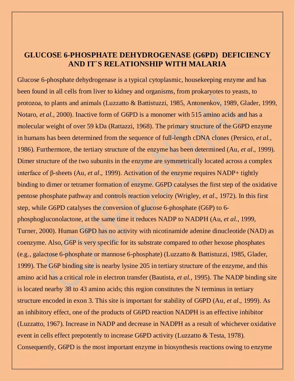
G6PD is the key enzyme in the oxidative pentose phosphate pathway. The first step of the
pentose phosphate pathway is catalyzed by G6PD. In this step, NADP+ is reduced to NADPH.
The second enzymatic step in thispathway is also associated with the reduction of NADP+ to
NADPH. The NADPHproduced as a consequence of these reactions reduces oxidized
glutathione (GSSG)to reduced glutathione (GSH) in a reaction catalyzed by glutathione reductase.GSH then reduces hydrogen peroxide, a powerful oxidant produced in the course of
cellular metabolism and as a consequence of the inflammatory response and other endogenous
and exogenous oxidants, in a reaction catalyzed by glu-tathione peroxidase (Newsholme and
Leech, 1983; Beutler, 1983; WHO WorkingGroup, 1989; Luzzatto and Mehta, 1989)., and
ribulose-5-phosphate,produced, a precursor of DNA, RNA, and ATP, emerge from G6P
(Turner, 2000). The most important reducing agent in the cytoplasm of cells is NADPH
(Koehler & Van Noorden, 2003). The second enzymatic step in this pathway is NADPH
production as a consequence of reactions that reduce oxidized glutathione (GSSG) to reduced
glutathione (GSH). The only defense against oxidant stress in the red blood cell (RBC) is GSH
production (Friedman, 1979, Group, 1989, Peters & Van Noorden, 2009).
In unstressed, normal erythrocytes, the G6PD activity is only about 2% of total capacity, which
is increased greatly to meet the challenge of an oxidant stress and GSH is maintained at stable
levels (Group, 1989, Peters & Van Noorden, 2009). The pentose phosphate pathway’s main
function is the generation of reducing capacity through the production of NADPH and
ultimately, GSH. This is essential for cell survival and is available in the erythrocyte for
generating reducing capacity (Greene, 1993).
CLASSIFICATION OF GLUCOSE-6-PHOSPHATE DEHYDROGENASE VARIANTS
More than 400 variants of G6PD have been distinguished based on their biochemical
characteristics, enzyme kinetics, physicochemical characteristics, and other parameters
(Luzzatto & Battistuzzi, 1985, Chen, et al., 1991, Greene, 1993). G6PD B+ is the most
commonly found enzyme type and it is used as a standard for normal enzyme activity and
electrophoretic mobility. For identification of other variants, G6PD B+ is used. The rate at
which NADP+ is reduced by glucose-6-phosphate with G6PD B+ as the catalyst is the standard
for activity. Based on this, enzyme activity relative to G6PD B+ variants are classified as fast,
normal, and slow in terms of electrophoretic mobility and as Classes I—V (Luzzatto, 1989,
Beutler, 1990, Greene, 1993, Segel, 2000, Betke K, Brewer GJ, Kirkman HN,
Luzzatto L,Motulsky AC, Ramot B, and Siniscalco M 1967). There are 5 classes for these
variants. Class I includes chronic nonspherocytic hemolytic anemia with a severe enzyme
deficiency (e.g., G6PD Minnesota, G6PD Tokyo, G6PD Campinas). Class II variants have
severe enzyme deficiency without chronic nonspherocytic hemolytic anemia (e.g., G6PD
mediterrian, G6PD Canton, G6PD Union, G6PD Kaiping). Class III variants includes medium
or mild enzyme deficiency, with the activity at 10-60% of G6PD B+ (e.g., G6PD Aˉ). Class IV
variants have a weak or no enzyme deficiency. The activity is 60-100 % of G6PD B+ (e.g.,
G6PD A+). Class V variants have increased enzyme activity (e.g., G6PD Hektoen) (Beutler,
1994, Segel, 2000).
GLUCOSE-6 PHOSPHATE DEHYDROGENASE DEFICIENCY AND MALARIA
As it is known, malaria is a parasitic disease that threatens 300-500 million people all over the
world. Malaria can be defined as the most deadly vector-borne disease in the world (Myrvang &
Godal, 2000). It is widespread in tropical and subtropical regions of Asia, Africa and the
American continents. Each year, malaria leads to deaths of millions of people all around the
world and a large percentage of deaths are seen in Sub-Saharan regions of Africa. The causative
agents of malaria are the Plasmodium parasites, which are transmitted to humans by the bites of
infected mosquitoes. If patients are not treated with antimalarial drugs, malaria can easily lead
to death. Five different types of Plasmodium species—P. falciparum, P. vivax, P. ovalae, P.
malariae and P.knowlesi—lead to this disease (Wernsdorfer & McGregor, 1988, Sutherland, et
al., 2010).
Plasmodium falciparum (P. falciparum) is the most serious and life-threatening form of the
disease. 80% of death cases are reported from patients that have been infected with P.
falciparum. It has also been demonstrated that resistance has been developed in this type of
parasites against current antimalarial drugs. It is generally seen in Africa, specifically in sub-
Saharan regions. Interestingly, falciparum-derived malaria cases have been recently reported in
various parts of the world where this parasite species was believed to be completely eradicated.
Plasmodium vivax (P. vivax) constitutes a milder form of the disease. Vivax infections generally
do not cause death. However, individuals that suffer from vivax infection also need to be
treated. Among all Plasmodium species, P. vivax is the one that shows the broadest geographic
distribution worldwide. Causative agents for 60% of malaria infections are reported as P. vivax
infections in India. This parasite has a liver stage and can remain in the body for years without
causing sickness. If the patient is not treated, the liver stage may re-activate and cause
relapses—malaria attacks—after months, or even years without symptoms.
Plasmodium ovale (P. ovale) is known as one of the other milder form of the disease. Like P.
vivax, it generally does not commonly lead to death. Nevertheless, infected individuals require
medical therapy. This parasite, similar to P.vivax, can live in the liver for long periods without
causing symptoms. Therefore, if it is not treated, reactivation of parasites can be observed in the
liver and this leads to relapse of the disease.
Plasmodium malariae (P. malariae) is also another milder form of the disease. It does not
commonly lead to death. However, it still requires treatment. This type of Plasmodium parasites
are reported to stay in the blood of some individuals for several decades.
Plasmodium knowlesi (P. knowlesi) causes malaria in macaques, but can also infect humans
(Mendis, et al., 2001, Singh, et al., 2004, Mueller, et al., 2007).
When life cycles of Plasmodium parasites are investigated, it is seen that the parasites multiply
in the liver of the human body, and then infect erythrocytes. Plasmodium parasites enter the
human body when bitten by an infective female
mosquito, which is called Anopheles. These mosquitoes become infected with malaria when
they take Plasmodium-containing blood from an infected person. Approximately one week
later, these parasites mix with the mosquito's saliva when the mosquito takes its next blood meal
from another person and this individual is injected with Plasmodium parasites when they are
being bitten (Bozdech, et al., 2003).
Multiplication of the parasites within erythrocytes enhances the severity of the disease and
cause symptoms such as anemia, fever, chills, nausea, flu-like illness, and, in severe cases,
coma, and death. Treatment of this disease can be achieved by using antimalarial drugs.
Primaquine, which is the most common antimalarial drug, can be used as a primary
prophylactic because it prevents primary parasitemia of Plasmodium species by destroying
these parasites in the liver before they reach the bloodstream and cause disease (Yazdani, et al.,
2006).
There is a strong relationship between malaria and G6PD deficiency diseases. In several
epidemiological studies, it was shown that distribution of malaria was nearly the same with
distribution of G6PD deficiency (Motulsky, 1961, Siniscalco & Bernini, 1961, Ganczakowski,
et al., 1995). This situation reveals two important facts. One of them is that G6PD deficiency
provides great protection from malaria, especially for falciparum infections. On the other hand,
using antimalarial drugs can cause life threatening hemolytic anemia in patients with G6PD
deficiency. Hence, malaria patients should be screened for their tendency to G6PD deficiency
before their treatment with antimalarial drugs.
When investigated in terms of cellular biology, Plasmodium parasite that
causes malaria use erythrocytes as host cells. Erythrocytes are also the most affected cells from
G6PD deficiency. This situation also suggests the relationship between the two diseases. In
several studies, it was demonstrated that G6PD deficiency provides a protection against malaria
infections. In one of the early studies, it was indicated that P. falciparum and P. vivax parasites
preferred to invade younger erythrocytes, which possessed high levels of G6PD enzyme. Since
enzyme levels are diminished in older erythrocytes, parasites do not prefer to invade these
erythrocytes. These studies suggested the protective effect of G6PD
deficiency from parasitemia (Allison & Clyde, 1961, Kruatrachue, et al., 1962). In the recent
past, Ruwando et al. also carried out a case-control study on more than 2,000 African children
and exhibited that risk of contracting malaria in patients that have the African form of G6PD
deficiency decreased at a rate of 46 to 58%. In this study, it was suggested that the selective
advantage of resistance to malaria was counterbalanced with selective disadvantageous results
of G6PD deficiency, and this stopped the rise of malaria frequencies in endemic regions
(Ruwende, et al., 1995). In another study, Ninokata et al. (2006) investigated 345 healthy adults
for G6PD deficiency on Phuket Island, which had been determined to be a malaria-endemic
region and found out that 10% of these individuals had G6PD deficiency. Interestingly, it was
observed that none of the individuals had molecular evidence of malaria infection. According to
this study, researchers postulated that G6PD deficiency provided an advantageous genetic trait
against malaria (Ninokata, et al., 2006).
The exact mechanism of this protection is still unknown. However there are two postulated
explanations. According to the first suggestion, it was found that parasites that cause malaria
can only survive in conditions with low oxygen levels (Clark, et al., 1989). This demonstrates
that these parasites are very susceptible to oxidative stress. It is known that in the pentose
phosphate pathway of erythrocytes, glucose-6 phosphate dehydrogenase (G6PD) enzyme has an
important role in production of NADPH and GSH. This is the only mechanism for erythrocytes
to survive. GSH that is produced by NADP+ reduction reacts with H2O2 and reduce it to H2O.
This prevents the generation of oxidative stress within red blood cells. Since oxidative stress is
the most important factor for the disruption of red blood cells, these cells are protected from this
effect. However, in G6PD deficient erythrocytes, G6PD activity is significantly reduced. In
G6PD A (-) variant, enzyme activity level reduces to 10 or 20% of normal levels, while enzyme
activity completely disappears in G6PD variant. Therefore, oxidative stress can be induced in
erythrocytes whose G6PD enzymes are deficient. In this situation, GSH is not produced and
H2O2 is not reduced to H2O and leads to oxidative stress. Hence, it is thought that since
malaria parasites are susceptible to oxidative stress, they do not live within the erythrocytes
where their maturation occurs (Toncheva & Tzoneva, 1985, Greene, 1993). Additionally,
during oxidative stress, the loss of
potassium from the cell and from the parasite can cause the death of the parasite (Friedman &
Trager, 1981).
According to the second suggestion, Plasmodium parasites oxidize NADPH and reduce the
level of reduced glutathione (GSH) in erythrocytes. In the situation of G6PD deficiency, this
effect becomes more severe and induces oxidative-induced damage within erythrocytes.
Moreover, Plasmodium parasites break down hemoglobin and release toxic components like
iron and these substances lead to hemolysis. Hence, the development rates of Plasmodium
parasites are diminished. Additionally, red blood cells that are affected by oxidative stress are
damaged and eliminated by the immune system via phagocytosis. This elimination decreases
the growth of parasites much more since it occurs during an early ring-stage of parasites’
maturation. Therefore, all of these data indicate that G6PD deficiency can provide protection
against malaria infections.
Considering the relationship between G6PD deficiency and plasmodium infections, research
has aimed to develop antimalarial drugs that decrease the level of GSH within erythrocytes and
then produce hydrogen peroxide and the other free radical species in order to enhance the
inhibition of Plasmodium species (Mehta, et al., 2000, Fortin, et al., 2002, Kwiatkowski, 2005,
Prchal & Gregg, 2005).
REFERENCE
(1967) Standardization of procedures for the study of glucose-6-phosphate dehydrogenase.
Report of a WHO Scientific Group. World Health Organ Tech Rep Ser Vol.366 (No.:1-53).
(Betke K, Brewer GJ, Kirkman HN, Luzzatto L,Motulsky AC, Ramot B, and Siniscalco M
Adam A (1961) Linkage between deficiency of glucose-6-phosphate dehydrogenase and colourblindness. Nature Vol.189(No.:686.
Download G6PD is a typical cytoplasmic
G6PD_is_a_typical_cytoplasmic.pdf (PDF, 449.94 KB)
Download PDF
Share this file on social networks
Link to this page
Permanent link
Use the permanent link to the download page to share your document on Facebook, Twitter, LinkedIn, or directly with a contact by e-Mail, Messenger, Whatsapp, Line..
Short link
Use the short link to share your document on Twitter or by text message (SMS)
HTML Code
Copy the following HTML code to share your document on a Website or Blog
QR Code to this page

This file has been shared publicly by a user of PDF Archive.
Document ID: 0000211422.