m140000 (PDF)
File information
This PDF 1.4 document has been generated by / PDFill: Free PDF Writer and Tools, and has been sent on pdf-archive.com on 27/07/2015 at 13:54, from IP address 103.58.x.x.
The current document download page has been viewed 657 times.
File size: 214.6 KB (11 pages).
Privacy: public file


File preview
Biojournal of Science and Technology
Research Article
Plasma cholesterol modulate functions of neutrophils in
streptozotocin-induced type 1 diabetic rats
AHM Nurun Nabi*, Mahfuzur Rahman, Laila N islam
Department of Biochemistry and Molecular Biology, University of Dhaka, Dhaka-1000, Bangladesh.
*Corresponding author
AHM Nurun Nabi
Professor, Department of Biochemistry and Molecular
Biology, University of Dhaka, Dhaka-1000, Bangladesh,
Phone: 880-2-29661900/Ext. 7660, E-mail:
nabi@du.ac.bd
Published: 28-05-2014
Biojournal of Science and Technology Vol.1:2014
Academic Editor: Editor-in-Chief
Received: 15-04-2014
Accepted: 10-05-2014
Article no: m140000
This is an Open Access article distributed under the terms of the Creative Commons Attribution License
(http://creativecommons.org/licenses/by/4.0 ), which permits unrestricted use, distribution, and
reproduction in any medium, provided the original work is properly cited.
Abstract
Objectives: Our previous study demonstrated neutrophil dysfunction in streptozotocin (STZ)-induced
diabetic rats. This study was aimed to investigate the biochemical indices such as plasma levels of
cholesterol, triglycerides, creatinine, urea, alanine transaminase and aspatate transaminase in diabetic and
control rats and thus, investigate their relationship with neutrophil functions. Methods: Diabetes was
induced in Long Evans rats by an intraperitoneal injection of citrate bu
buffer
ffer dissolved streptozotocin
(STZ). Age matched control rats were injected with citrate buffer only. Neutrophils were isolated from
blood using standard dextran sedimentation followed by Ficoll-Hypaque centrifugation; morphological
changes in neutrophils, their ability to reduce nitroblue tetrazolium (NBT) dye and phagocytic activity
from both the groups of rats were evaluated formerly. Biochemical indices were measured by standard
colorimetric methods. Results: The average levels of glucose, triglycerides
triglycerides,, cholesterol, creatinine, urea
in the plasma of diabetic and control rats were 302.6 ± 87.5 vs 100.7 ± 11.5 mg/dL, 174.9 ± 18.6 vs 82.2
± 10.2 mg/dL; 250.8 ± 22.3 vs 165.2 ± 24.1 mg/dL; 0.94 ± 0.19 vs 0.81 ± 0.05 mg/dL; 77.1 ± 9.7 vs 26.8
v 5.8 mg/dL, respectively. The mean values of aspartate transaminase (AST) and alanine transaminase
(ALT) in diabetic and control rats were 141.4 ± 28.0 vs 61.6 ± 18.6 IU/L and 61.4 ± 13.6 vs 48.5 ± 6.0
IU/L, respectively. Biochemical parameters measured in diabetic rats varied significantly (p < 0.001)
compared to those of control rats. Plasma indices such as triglycerides, cholesterol, creatinine, urea, AST
and ALT had no relation with the functions of neutrophils. However, multidimensional scaling found a
close relation between plasma cholesterol and phagocytic activity of neutrophils from diabetic rats.
Ability to reduce NBT dye was closely related to the morphology of the activated neutrophils. On the
other hand, levels of plasma glucose were distantly related to the ffunctions
unctions of neutrophils. Conclusion:
Thus, important liver and kidney functions indices, lipid profile parameters were significantly altered in
diabetic rats and plasma cholesterol modulated the phagocytic activity of neutrophils.
Keywords: Diabetic rats, phagocytic activity, plasma cholesterol, STZ-diabetes, neutrophils.
ISSN 2410-9754
Vol:1, 2014
INTRODUCTION
Neutrophils, part of the innate immune response,
are the first line of defense in hosts. Numerous
studies have demonstrated altered neutrophil
functions in diabetes and it is suggested that the
impaired neutrophil functions [e.g., chemotaxis,
phagocytosis, nitroblue tetrazolium (NBT) dye
reduction ability etc.] cause the susceptibility to
infections in diabetics (Coopan, 1985; Reeves and
Wilson, 1992). The chemotactic activity of
neutrophils from diabetic patients is significantly
lower compared to those of healthy controls
(Mowat and Baum, 1971). Studies of the
phagocytic and microbicidal activities of
neutrophils from diabetic patients reveal, with few
contrasting results, an impairment of these
functions. Decreased bactericidal activity (Tan et
al, 1975), impairment of phagocytosis and
decreased release of lysosomal enzymes (Bagdade
et al, 1972), and reduced production of reactive
oxygen species (Nielson and Hindson, 1989) by
neutrophils of diabetic patients have been
described. Furthermore, reduction in leukocyte
phagocytosis and bactericidal activity showed a
significant correlation with increases in blood
glucose levels (Jakelic et al, 1995).
Hyperglycemia, one of the characteristic
manifestations of diabetes mellitus, has been found
to be associated with neutrophil dysfunctions
(Nabi et al, 2005). Formation of advanced
glycation end products (AGEs) through an
interaction between glucose and lipids and/or
proteins is one of the causes of long-term
complications in diabetes (Brownlee, 2001).
Glycosylated proteins isolated from the serum of
diabetic rats affect membrane permeability and
migration, reduce the rolling and adhesion abilities
of leukocytes in alloxan-induced diabetic rats
(Sannomiya et al., 1997; Masuda et al., 1990).
Further, AGEs in human is linked to a rise in
@2014, GNP
intracellular Ca2+ and to actin polymerization
(Collison et al., 2002). Actin polymerization in
neutrophil plays important role in chemotactic
action of neutrophils that is crucial to exhibit its
normal functions. A positive association between
polyol pathway activation and leukocyte
dysfunction in experimental diabetes mellitus has
been reported and hypothesized that the
accelerated formation of sorbitol in diabetic
animals may increase the intracellular osmolarity
or decrease the availability of the enzyme co-factor
NADPH, leading to a disturbance of endothelial
cell
functions
that
might
alter
leukocyte-endothelial cell interactions (Cruz et al.,
2000).
Epidemiological studies have established a strong
correlation between elevated total cholesterol
levels in serum and morbidity and mortality from
myocardial infarction (Thomas et al, 1993).
Elevated numbers of circulating neutrophils have
been shown to be predictive of cardiovascular
events independent of serum cholesterol levels
(Guasti et al, 2011) In a more recent study, a direct
mechanistic link between hypercholesterolemia
and proliferation of myeloid progenitor cells and,
hence, neutrophilia and monocytosis has been
identified (Murphy et al, 2011 and Weber et al,
2011). High dose of Statin, a cholesterol lowering
agent, has been demonstrated to lower the
migration ability of neutrophils significantly in
healthy volunteers (Kinsella et al, 2011). Further,
free cholesterol has been found to be associated
with the altered lipid raft structure of cell
membrane and function regulating neutrophil ca2+
entry and respiratory burst (Kolenkode et al, 2007).
Our previous study demonstrated neutrophil
dysfunction in streptozotocin (STZ)-induced
diabetic rats (Nabi et al., 2005). This study was
aimed to investigate the biochemical indices such
as plasma levels of cholesterol, triglycerides,
Biojournal of Science and Technology
Page |1
ISSN 2410-9754
Vol:1, 2014
creatinine, urea, alanine transaminase and aspartate
transaminase in diabetic and control rats and thus,
investigate their relationship with the previously
studied neutrophil functions.
MATERIALS AND METHODS
Preparation for streptozotocin-induced type 1
diabetes mellitus in the rats and collection of
blood and plasma
A total of 30 Long Evan rats (Male: 15; Female: 15)
each 2 weeks of age were kept in the plastic cages
with even floors covered with wood shavings in
the animal house of the department of
Biochemistry and Molecular Biology, University
of Dhaka, Bangladesh. The initial average body
weight of the male rats was 145.3 ± 5.6 gm and of
the female rats was 140.85 ± 4.3 gm. These
animals were kept under constant temperature with
a 14 hour light and 10 hour dark cycle. About 5-6
gm of balanced pelleted rat food was supplied
thrice a day. These animals had also free access to
drinking water. These conditions were maintained
for the next 4 weeks.
Protocol
for
the
preparation
of
streptozotocin-induced type 1 diabetic rats has
been described in our previous study (Nabi et al.,
2005) using intraperitoneal injection of
streptozotocin (STZ) dissolved in citrate buffer (65
mg/Kg body weight) at the age of 4 weeks (185.5
± 10.2 gm and 180.7 ± 11.2 gm body weights for
10 male and 10 female rats, respectively). Age
matched control rats (5 male and 5 female) were
injected with citrate buffer only. After
anesthetizing in a chamber containing diethyl ether,
blood samples were collected by sacrificing each
diabetic and control rats into a heparin-containing
falcon tube. Immediately after collection, 2.0 ml of
blood was transferred into fresh tube and
centrifuged at 3000 rpm for 10 minutes. The
@2014, GNP
plasma was collected and stored at –20OC until
further analysis.
Isolation of neutrophils, polarization assay and
NBT dye reduction tests
Neutrophils were isolated from the freshly
collected blood samples of control and diabetic
rats by dextran (Sigma) sedimentation followed by
centrifugation on Ficoll-Hypaue (Pharmacia,
Uppsala, Sweden) as described elsewhere (Islam
and Nabi, 2003; Nabi et al, 2005). The separated
neutrophils were resuspended in a minimal volume
(1.0 – 1.5 ml) of Hank's Balanced Salt Solution
mixed with 3-(N-morpholino)-propanesulfonic
acid (HBSS-MOPS). The percentage of polarized
cells was assessed by viewing the cell preparation
under Light microscope using a 40X objective
(Nabi and Islam, 2001; Islam and Nabi, 2003; Nabi
et al, 2005). At least 300 cells were counted from
each preparation. NBT dye reduction test was
performed according to the protocol described
earlier (Nabi and Islam, 2001; Nabi et al, 2005).
Briefly, neutrophils (2 x 10⁶ cells/mL) treated with
yeast activated serum, incubated for 30 minutes at
37OC and then, aliquots of NBT (Sigma) solution
was added into the cells and incubated for 1 hour
at 37OC. The unused NBT was removed through
washing and the reduced dye was extracted in
dioxan (Sigma) and quantitated at 520 nm.
Biochemical analyses of the plasma samples
Total plasma cholesterol, triglycerides, plasma
creatinine, urea and activities of plasma AST and
ALT were measured by standard colorimetric
method in an Autoanalyzer (UK) using kits
purchased from RANDOX, UK. Briefly, plasma
cholesterol was measured by oxidation of
cholesterol using cholesterol oxidase, triglycerides
by enzymatic hydrolysis using lipases, creatinine
by Jaffe method using alkaline picrate, urea using
urease method, ALT and AST by kinetic methods
Biojournal of Science and Technology
Page |2
ISSN 2410-9754
Vol:1, 2014
using lactate dehydrogenase and NADH.By means
of respective units, results of each parameter were
expressed.
Multi dimensional scaling or Euclidean distance
model was performed using SPSS package.
Phagocytosis assay
Baker’s yeasts (Saccaromyces cerevisiae; Gist
brocades, Holland) were preincubated with fresh
control serum for opsonization to perform
phagocytosis assay. Neutrophils (1 x 10⁶cells/mL,
from control and diabetic rats) were taken onto
clean glass slides and incubated for 5 minutes at
37OC. A few drops of prepared yeasts at 1 x
108/mL were then added to the neutrophils and
incubated for a further 5 minutes at 37OC. The
scoring was done according to our previous
protocol (Islam and Nabi, 2003; Nabi et al, 2005).
The percentage of phagocytic cells and the number
of yeast cells attached per 100 randomly chosen
neutrophils were counted by examining at least
300 neutrophils from each preparation (controls
and diabetics) under the oil immersion lens.
RESULTS
Biochemical analyses
The development of diabetes was confirmed by the
presence of hyperglycemia (blood glucose level >
230 mg/dL) as described previously (Nabi et al,
2005). Plasma glucose levels were determined by
the glucose oxidase method using blood samples
obtained from the animal tail. The rats were used
for the experiments 1 week after receiving STZ
injection. The average levels of glucose in the
plasma of diabetic and control rats were 302.6 ±
87.5 and 100.7 ± 11.5 mg/dL, respectively. Other
biochemical indices examined from the plasma of
both the groups of rats have been presented in
Table 1 and shown in Figure 1. In diabetic and
control rats, the average levels of plasma
triglycerides were 174.9 ± 18.6 vs 82.2 ± 10.2
Statistical analyses
mg/dL; total cholesterol were 250.8 ± 22.3 vs
The results were expressed as mean ± SD. To
165.2 ± 24.1 mg/dL; plasma creatinine were 0.94 ±
compare the differences between neutrophils from
0.19 vs 0.81 ± 0.05 mg/dL; plasma urea were 77.1
the control and diabetic rats, independent Student's
± 9.7 vs 26.8 v 5.8 mg/dL, respectively. The mean
values of aspartate transaminase and alanine
t-test was performed. Correlation was determined
transaminase in diabetic and control rats were
by using non-parametric Spearman's rho test. A p
141.4 ± 28.0 vs 61.6 ± 18.6 IU/L and 61.4 ± 13.6
value of less than 0.05 was considered significant.
vs 48.5 ± 6.0 IU/L, respectively.
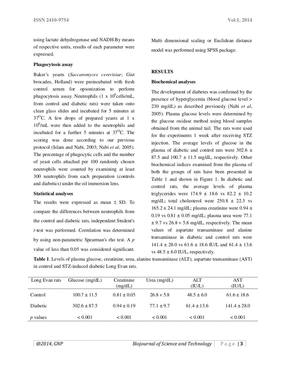
Table 1. Levels of plasma glucose, creatinine, urea, alanine transaminase (ALT), aspartate transaminase (AST)
in control and STZ-induced diabetic Long Evan rats.
Long Evan rats
Glucose (mg/dL)
Creatinine
(mg/dL)
Urea (mg/dL)
ALT
(IU/L)
AST
(IU/L)
Control
100.7 ± 11.5
0.81 ± 0.05
26.8 v 5.8
48.5 ± 6.0
61.6 ± 18.6
Diabetic
302.6 ± 87.5
0.94 ± 0.19
77.1 ± 9.7
61.4 ± 13.6
141.4 ± 28.0
p values
< 0.001
< 0.001
< 0.001
< 0.001
< 0.001
@2014, GNP
Biojournal of Science and Technology
Page |3
ISSN 2410-9754
Vol:1, 2014
P<0.001
P<0.001
210
190
LDL concentration (mg/dL)
Cholesterol concentration (mg/dL)
350
300
250
200
150
170
150
130
110
90
70
100
50
Controls
Diabetics
Controls
Diabetics
Figure 1. Levels of cholesterol (A) and triglycerides (B) in the plasma of diabetic and control rats. The mean
levels of plasma triglycerides were 174.9 ± 18.6 and 82.2 ± 10.2 mg/dL in diabetic and control rats,
respectively that varied significantly (p < 0.001).
that the ability to reduce nitroblue terazolium dye
Statistical analysis using student’s t test showed an
by the neutrophils (e) of the study rats was closely
increase levels of all the biochemical parameters
related to the percentages of the polarized
that measured in diabetic rats varied significantly
neutrophils (f).
(p < 0.001) compared to those of control rats
(Table 1 and Figure 1).
Association of biochemical indices with the
phagocytic activity of neutrophils
Association of biochemical indices with
According to our previous study, it was perceived
polarization of neutrophils and NBT dye
that neutrophils from the diabetic rats were
reduction
statistically less phagocytic than those from the
Our previous reports revealed that at the base line
control rats (61 ± 7% vs 87 ± 4%, respectively; p <
level, neutrophils from diabetic rats were
0.001) by phagocytosing significantly (p < 0.001)
significantly more polarized (p < 0.001) compared
less number of opsonized yeast particles (282 ± 16)
to those from control rats (30 ± 4 vs 13 ± 3) (Nabi
compared to those of the control cells (381 ± 17)
et al, 2005). We also showed that neutrophils from
(Nabi et al, 2005). Though our previous findings
diabetic rats could reduce significantly more NBT
demonstrated that percentages of phagocytic
dye (0.12 ± 0.03 vs 0.04 ± 0.01, respectively; p <
neutrophils as well as efficiency of the neutrophils
0.001) than those from control rats at the baseline
i.e., number of yeast particles phagocytosed by the
level (Nabi et al, 2005). These functions exhibited
cells were inversely related to the levels of plasma
by diabetic and control neutrophils did show any
glucose in diabetic rats (Nabi et al, 2005), we did
relationship with the biochemical indices
not find any relationship between the immune
examined (Table 2 and 3). However, Euclidean
function mediated by the neutrophils and plasma
distance model represented in Figure 2 indicated
cholesterol/triglycerides
using
bivariate
@2014, GNP
Biojournal of Science and Technology
Page |4
ISSN 2410-9754
Vol:1, 2014
Spearman’s rho analysis in case of neither diabetic
nor control rats (Table 2 and 3). Interestingly,
multidimensional scaling plot demonstrated a close
relation between the levels of plasma total
cholesterol and the percentages of phagocytic
neutrophils as shown in Figure 2.
2.0
a
1.5
1.0
fe
.5
c
Dimension 2
0.0
-.5
b
-1.0
d
-1.5
-2.0
-1.5
-1.0
-.5
0.0
.5
1.0
1.5
Dimension 1
Figure 2. Euclidean distance model or multiple distance scaling indicated that the ability to reduce nitroblue
terazolium dye by the neutrophils (e) of the study rats was closely related to the percentages of the polarized
neutrophils (f). On the other hand, phagocytic activity of the neutrophils (b) was closely related to that of the
plasma endogenous cholesterol (d). This model further showed that plasma glucose (a) and total number of
yeasts phagocytosed by neutrophils (c) are distantly related.
DISCUSSION
The levels of biochemical indices such as
important metabolites of lipid profile panel e.g.,
plasma cholesterol, triglycerides; of kidney
function e.g., creatinine and urea; of liver function
activities of serum enzymes such as aspartate
transaminase and alanine transaminase were
studied in streptozotocine-induced type 1 diabetic
rats and thus, association of these indices with the
possible alteration of the functions of neutrophils
were investigated in this study. Plasma ALT and
AST levels were measured to evaluate the hepatic
functions. This study reveals statistically
@2014, GNP
significant (p < 0.001) increased levels of AST and
ALT in the plasma of diabetic rats compared to
that of control rats (Table 1). The increase in
aminotransferases levels may be due to the cellular
damage in the liver caused by STZ-induced
diabetes. Thus, our result supports the data of other
researchers who found elevated levels of ALT and
AST in diabetic rats (Zafar et al, 2009; Baxter and
Schofield, 1980). Voss et al. (1988) also reported
similar finding by proposing time dependent rise in
AST, ALT, and alkaline phosphates (ALP) levels in
STZ-induced diabetic rats. Moreover, we also
found that levels of creatinine and urea were
Biojournal of Science and Technology
Page |5
ISSN 2410-9754
Vol:1, 2014
significantly elevated in the plasma of diabetic rats
(Table 1) which matched with the data reported by
Voss et al. (1980). Thus, this data indicated that
normal metabolic processes are altered in
STZ-induced diabetic rats by affecting both liver
and kidneys.
Table 2. Correlation of the plasma glucose, triglycerides, total cholesterol, creatinine and urea
with functional indices of neutrophils such as percent polarized, percentages of phagocytic
neutrophils, total number of opsonized yeasts phagocytosed and ability to reduce NBT dye by
neutrophils from diabetic rats.
Glucos
e
(mg/d
L)
Glucose
(mg/dL)
Creatinine
(mg/dL)
Urea
(mg/dL)
%Phagocyti
c cells
Creatini
ne
(mg/dL)
Urea
(mg/d
L)
%Phagoc
ytic cells
Total
number of
yeast
phagocyto
sed
NBT
reducti
on
%polari
zed cells
Plasma
triglycer
ide
(mg/dL)
1.000
0.133
1.000
0.170
-0.081
1.000
-0.497
*a
0.013
-0.406
1.000
-0.500
*a
0.264
-0.230
0.245
1.000
NBT
0.044
-0.132
0.111
reduction
%polarized
-0.193
0.007
-0.034
cells
Plasma
triglyceride -0.175
-0.012 0.449*
(mg/dL)
Plasma
cholesterol
0.185
-0.429
-0.213
(mg/dL)
*Correlation is significant at the 0.05 level
(2-tailed).
a
Nabi et al, 2005.
0.132
-0.179
1.000
0.343
0.113
-0.230
1.000
0.163
-0.026
0.003
0.172
1.000
0.067
-0.193
0.225
0.030
-0.017
Total
number of
opsonized
yeast
phagocytos
ed
@2014, GNP
Plasma
choleste
rol
(mg/dL)
Biojournal of Science and Technology
1.000
Page |6
ISSN 2410-9754
Vol:1, 2014
Table 3. Correlation of the plasma glucose, triglycerides, total cholesterol, creatinine and urea with functional
indices of neutrophils such as percent polarized, percentages of phagocytic neutrophils, total number of
opsonized yeasts phagocytosed and ability to reduce NBT dye by neutrophils from control rats.
Glucos
e
(mg/dL
)
Glucose
(mg/dL)
Plasma
triglycerid
e (mg/dL)
Plasma
cholestero
l (mg/dL)
Creatinin
e
(mg/dL)
Urea
(mg/dL
)
%Phagocyti
c cells
Total
number of
yeast
phagocytose
d
NBT
reductio
n
%polarize
d cells
1.000
Plasma
triglyceride
(mg/dL)
0.078
1.000
Plasma
cholesterol
(mg/dL)
0.267
0.237
1.000
Creatinine
(mg/dL)
-0.237
-0.585
0.061
1.000
Urea
(mg/dL)
-0.284
0.030
0.607
0.469
1.000
%Phagocyti
c cells
-0.103
-0.201
-0.568
0.116
-0.390
1.000
Total
number of
opsonized
yeast
phagocytose
d
0.187
-0.369
-0.158
0.048
-0.296
0.024
1.000
NBT
reduction
0.466
-0.090
0.413
0.012
0.163
0.310
-0.248
1.000
%polarized
cells
0.613
0.328
0.103
-0.733*
-0.492
-0.397
0.200
0.042
1.000
*Correlation is significant at the 0.05 level
(2-tailed).
In vitro animal and clinical research indicated
extensive relationship between serum lipids and
the immune system (Sullivan, 1994; Kelley and
Bendich, 1996). From the basic metabolic
standpoint of view, we know that lipoprotein
particles deliver cholesterol, various phospholipids
and fat soluble antioxidants to cells which are
essential for maintaining the integrity of cell
membrane and optimal immune function thorugh
the production of eicosanoid and anti-oxidants
@2014, GNP
(Heiniger et al, 1978; Pace and Eshfahani, 1987;
Bendich, 1990). Antigen presenting activity of
monocytes and chemotactic activity of some cell
lines can be increased by cholesterol (Hughes et al,
1992; Kreuzer et al, 1991). Another study reported
that diet-induced hypercholesterolemia in animals
reduce macrophage response and phagocytic
function (Kos et al, 1979).
Cholesterol is present in human plasma at
Biojournal of Science and Technology
Page |7
ISSN 2410-9754
Vol:1, 2014
concentrations of milligrams per milliliter. The
vast majority is complexed in lipoproteins.
However, although clinical free cholesterol is
albumin-bound
and
albumin
can
delay
cholesterol’s effects on signaling in vitro, cells are
continuously exposed to cholesterol in vivo in
equilibrium where cholesterol desorbs from
albumin to cell membranes. Moreover, albumin
concentrations vary markedly from plasma to the
interstitium in health, and may vary further as a
function of disease states. It is reported that high
serum or tissue-free cholesterol availability could
activate immune cells that contribute to the
progression of atherosclerotic lesions (Kolenkode
et al, 2007). With respect to lipid effects on
neutrophil phagocytosis, the phagocytic capacity in
most human and animal studies appears to be
decreased in vitro by lower chain triglycerides or
LCT (Wiernik et al, 1983; Usmani et al, 1988;
Rasmussen et al, 1988). Also, depressed
phagocytosis, oxygen radical production and
Fc-receptor expression have been observed after
exposure of neutrophils to LCT (Cleary and
Pickering, 1983).
The earliest morphological response of leucocytes
to a chemoattractant is a change from a spherical
to a polarized shape with formation of an extended,
ruffled, anterior veil or lamellipodium. This
polarization is accompanied by functional
polarization, since the lamellipodium remains
organelle-free, is rich in filamentous actin, and
several cell surface proteins have been reported to
become concentrated at the front of the cell. In
various leucocytes these proteins include Fc
receptors (Walter et al., 1980; Wilkinson et al.,
1980), Thy-1 (Shields and Haston, 1985), CD15
and CD45 (Haston and Maggs, 1990), and
urokinase-type plasminogen activator receptor
(Estreicher et al. 1990). In the present study, it has
been demonstrated through multi-dimensional
@2014, GNP
scaling that phagocytic activity of the neutrophils
are closely related to that of the levels of plasma
cholesterol (Figure 2). Thus, it is possible that the
equilibrium
between
the
albumin-bound
cholesterol and membrane cholesterol has been
disturbed which might have altered baseline
morphology of neutrophils followed by change in
the normal distribution of receptors responsible for
phagocytosis as reported by Cleary and Pickering
(1983). This has also been reflected by the
significantly lower percentages of phagocytosis
mediated by diabetic neutrophils. Previous
findings revealed enhanced phagocytic activity by
the activated neutrophils (Kozel et al, 1987). Thus,
it was expected that activated diabetic neutrophils
at the baseline level should demonstrate higher
phagocytic
activity.
However,
decreased
phagocytic activity of the diabetic neutrophils as
well as their decreased efficiency and its close
association with plasma cholesterol might be
attributed to the possible i) shedding of the
receptors and/or ii) sequestration of the receptors
and/or iii) alteration of membrane-bound receptor
mediated cell signaling required to maintain
optimal phagocytic activity. Thus, endogenous
plasma cholesterol elevated due to type 1 diabetes
in STZ-induced diabetic rats may alter the
functions of neutrophils in vitro. The short coming
of the current study is that we could not evaluate
the effects of exogenous cholesterol on the
neutrophil functions. However, future research
work should be performed to observe the
dose-dependent and time-dependent effects of
cholesterol on neutrophil functions, status of
different functional receptors on the plasma
membrane as well as on the cell signaling
mechanism.
Biojournal of Science and Technology
Page |8
Download m140000
m140000.pdf (PDF, 214.6 KB)
Download PDF
Share this file on social networks
Link to this page
Permanent link
Use the permanent link to the download page to share your document on Facebook, Twitter, LinkedIn, or directly with a contact by e-Mail, Messenger, Whatsapp, Line..
Short link
Use the short link to share your document on Twitter or by text message (SMS)
HTML Code
Copy the following HTML code to share your document on a Website or Blog
QR Code to this page

This file has been shared publicly by a user of PDF Archive.
Document ID: 0000291800.