Project 2 TMJ TJR Group 4 (PDF)
File information
This PDF 1.5 document has been generated by LaTeX with hyperref package / pdfTeX-1.40.16, and has been sent on pdf-archive.com on 27/03/2016 at 00:55, from IP address 98.248.x.x.
The current document download page has been viewed 751 times.
File size: 472 KB (35 pages).
Privacy: public file


File preview
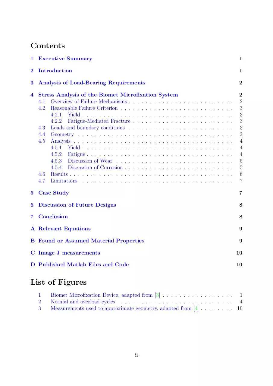
BIOE/ME C117 Project II - Group 4
Total Temporomandibular Joint Replacement
Leo Brossollet, Kristy Ip, Lulu Li, Josephine Wu
March 17, 2016
i
Contents
1 Executive Summary
1
2 Introduction
1
3 Analysis of Load-Bearing Requirements
2
4 Stress Analysis of the Biomet Microfixation
4.1 Overview of Failure Mechanisms . . . . . . .
4.2 Reasonable Failure Criterion . . . . . . . . .
4.2.1 Yield . . . . . . . . . . . . . . . . . .
4.2.2 Fatigue-Mediated Fracture . . . . . .
4.3 Loads and boundary conditions . . . . . . .
4.4 Geometry . . . . . . . . . . . . . . . . . . .
4.5 Analysis . . . . . . . . . . . . . . . . . . . .
4.5.1 Yield . . . . . . . . . . . . . . . . . .
4.5.2 Fatigue . . . . . . . . . . . . . . . . .
4.5.3 Discussion of Wear . . . . . . . . . .
4.5.4 Discussion of Corrosion . . . . . . . .
4.6 Results . . . . . . . . . . . . . . . . . . . . .
4.7 Limitations . . . . . . . . . . . . . . . . . .
System
. . . . . .
. . . . . .
. . . . . .
. . . . . .
. . . . . .
. . . . . .
. . . . . .
. . . . . .
. . . . . .
. . . . . .
. . . . . .
. . . . . .
. . . . . .
.
.
.
.
.
.
.
.
.
.
.
.
.
.
.
.
.
.
.
.
.
.
.
.
.
.
.
.
.
.
.
.
.
.
.
.
.
.
.
.
.
.
.
.
.
.
.
.
.
.
.
.
.
.
.
.
.
.
.
.
.
.
.
.
.
.
.
.
.
.
.
.
.
.
.
.
.
.
.
.
.
.
.
.
.
.
.
.
.
.
.
.
.
.
.
.
.
.
.
.
.
.
.
.
.
.
.
.
.
.
.
.
.
.
.
.
.
.
.
.
.
.
.
.
.
.
.
.
.
.
.
.
.
.
.
.
.
.
.
.
.
.
.
.
.
.
.
.
.
.
.
.
.
.
.
.
2
2
3
3
3
3
3
4
4
4
5
5
6
7
5 Case Study
7
6 Discussion of Future Designs
8
7 Conclusion
8
A Relevant Equations
9
B Found or Assumed Material Properties
9
C Image J measurements
10
D Published Matlab Files and Code
10
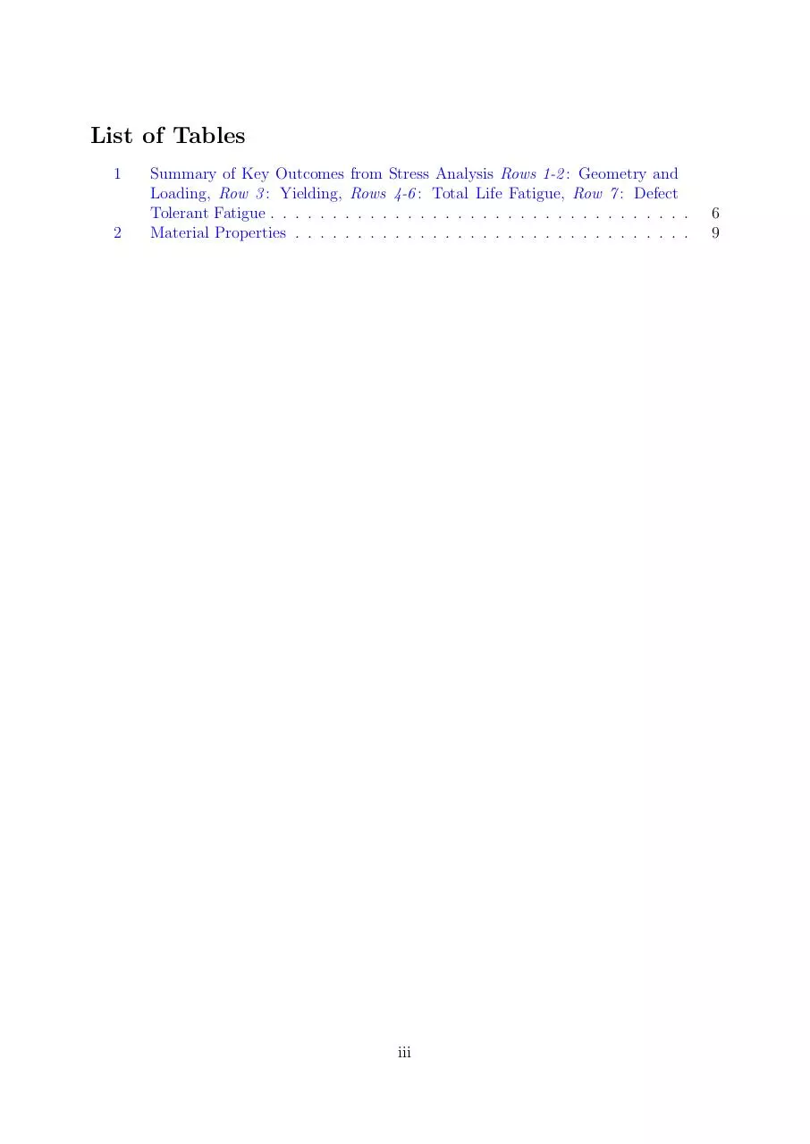
List of Figures
1
2
3
Biomet Microfixation Device, adapted from [3] . . . . . . . . . . . . . . . . . 1
Normal and overload cycles . . . . . . . . . . . . . . . . . . . . . . . . . . . 4
Measurements used to approximate geometry, adapted from [4] . . . . . . . . 10
ii
List of Tables
1
2
Summary of Key Outcomes from Stress Analysis Rows 1-2 : Geometry and
Loading, Row 3 : Yielding, Rows 4-6 : Total Life Fatigue, Row 7 : Defect
Tolerant Fatigue . . . . . . . . . . . . . . . . . . . . . . . . . . . . . . . . . .
Material Properties . . . . . . . . . . . . . . . . . . . . . . . . . . . . . . . .
iii
6
9
1
Executive Summary
Total temporomandibular joint (TMJ) replacement is a last resort treatment for progressive TMJ disorders. TMJ replacement presents unique challenges, as the joint is bicondylar, small, and anatomically complex. Of three major devices available for TMJ
replacement, we chose to perform a stress analysis of the Biomet Microfixation system,
examining yield, fatigue, wear, and corrosion as potential modes of failure. It was concluded that while the device is safe from purely mechanically failure, it is highly susceptible
to mechanically-induced biological failure such as inflammatory responses from wear and
corrosion debris.
2
Introduction
Temporomandibular joint (TMJ) disorders are uniquely difficult to treat due to the
small size and anatomical complexity of the joint [1]. The TMJ is a bicondylar joint, where
mechanical changes on one side affect the other [2]. Patients with TMJ disorders tend
to initially opt for non-surgical treatments, but because of the progressive nature of the
condition, about 1% of patients may eventually choose surgical treatments [1].
The most extreme and invasive of surgical options is total joint arthroplasty, or TMJ
replacement, and has success rates ranging from 30-100% [2]. The prosthesis includes a
temporal component, which replaces the glenoid fossa and interpositional disk to function
as an articular bearing. The device also has a mandibular component, which replaces the
condyle as a counter-bearing [1].
The Biomet Microfixation TMJ implant is one of three major systems available for TMJ replacement, and it is the
device we will be analyzing [Figure 1]. It
can be used for either unilateral or bilateral
joint reconstruction. The temporal component of the system, also known as the fossa
prosthesis, is fixed to the base of the skull.
Made entirely of ultra high molecular weight
polyethylene (UHMWPE), the fossa prosthesis is about 25mm across and available
in small, medium, or large sizes [3, 4]. The
mandibular component of the system, also
known as the condylar prosthesis, is fixed
Figure 1: Biomet Microfixation Device,
to the ramus. The component is primarily
adapted from [3]
made of cobalt-chrome (CoCr) alloy and its
underside is coated with a titanium plasma spray. The Biomet mandibular component is
45, 50, or 55mm long, paired with standard, offset, or narrow styles. Both the fossa and
condylar components are fixed with titanium screws [3].
In this report, we examine the load-bearing requirements of TMJ replacement systems, review relevant literature on failure modes, perform a stress analysis of the Biomet
Microfixation TMJ TJR system, and conclude with a discussion of implications for future
1
designs.
3
Analysis of Load-Bearing Requirements
The prosthesis must restore two primary functions of the joint: speech and mastication.
For both actions, the joint must have at least 50% of healthy translational and rotational
kinematics. There should be medial-lateral and anterior-posterior translation, and sagittal
and transverse plane rotation of the jaw. The TMJ is subjected to primarily compressive
and also shear loads under cyclic loading. Due to incongruent anatomy of the joint, the
contact area is quite small and experiences large stresses during loading [1]. In mastication,
incisors transfer the most force to the TMJ (60-90%), whereas molars transfer the least
(5-70%) [1, 5]. A TMJ replacement should be able to sustain 800N of compressive loads
and 200N of shear loads for 400,000 cycles per year. As many patients who undergo TMJ
replacement are in their 30s, the device should have a lifetime of at least 20 years [1].
This project examines possible failure mechanisms under normal loading conditions in the
Biomet TMJ replacement system.
4
4.1
Stress Analysis of the Biomet Microfixation System
Overview of Failure Mechanisms
Failure of TMJ total joint replacement (TJR) devices typically manifests as pain around
the joint space or a decrease in jaw mobility. Of the three devices on the market today,
there is not a significant depth of literature concerning the underlying mechanical failure
modes. It is reasonable to conclude that most TMJ TJR devices on the market today do
not fail due to yielding, fracture, or bending, but from wear, corrosion, infection, and other
biological responses. These latter failures are difficult to predict quantitatively.
In a study of 442 Biomet implants in 228 patients, removals of devices or revision
surgeries were prompted only by infection or bone formation around the joint space that
limited opening of the jaw [6]. While this was only a 3-year follow-up, other studies indicate
that revision surgeries with longer follow-up periods were caused by similar bone formation
[7, 8]. A 2007 follow-up of patients with custom TMJ Concepts devices revealed that
85% reported an increase in quality of life at 10 years [9]. The overall clinical success
and lack of revision surgery would suggest that these devices do not undergo mechanical
failure within 10 years. Another study looked at six patients that presented with symptoms
associated with fibrous capsule formation around the joint. Only one case of six presented
with inflammatory reactions in surrounding tissue, and none featured foreign body particles
in tissue surrounding the joint space [4]. In a study of 28 failed retrieved Christensen
implants, wear characteristics were observed and discussed, but the failure criteria and
modes were not explicitly defined [10]. As there is not a consensus in medical literature
suggesting preference for a specific failure mode, we will examine the Biomet Microfixation
device for failure in yield, fatigue, wear, and corrosion.
2
4.2
4.2.1
Reasonable Failure Criterion
Yield
UHMWPE and CoCr are both ductile materials, as they yield before fracture. As
yielding causes plastic deformation in both materials, yielding in either could change the
geometry of the TJR system and result in significant discomfort or pain. Therefore we treat
yielding of either the UHMWPE or CoCr as failure. However, UHMWPE has a lower elastic
modulus and compressive strength, therefore we use a von Mises yielding criterion for the
UHMWPE to evaluate safety against yielding in the system. As yielding would precede fast
fracture from a single load cycle in this system, we next consider fatigue-mediated fracture.
4.2.2
Fatigue-Mediated Fracture
A total-life philosophy of fatigue and Miner’s rule are used to determine damage incurred in the device with daily use. Additionally, a defect-tolerant, crack-propagation
scheme is used to evaluate device susceptibility to crack growth and fast fracture. Although susceptibility to failure by wear is difficult to quantify, we recognize and discuss
qualitative factors that affect wear mechanisms, including the Hertz contact stresses and
frictional forces at the contact surface that may liberate debris. Likewise, corrosion resistance is assessed in terms of material properties of the components, device construction,
and implementation. Material properties for UHMWPE and CoCr are listed in 2.
4.3
Loads and boundary conditions
Two loading conditions are considered in the stress analyses: maximum bite force
(MBF, 800N) and chewing (mastication, 20-400N, 2000 cycles/day for 20 years) [1, 2]. The
resultant contact forces on the TMJ can be estimated to be at most 70% of the magnitude of
molar forces; load transfer ratios differ along the mandible and vary based on the dentition
and anatomy of each patient [1]. The joint reaction force is further simplified to be a point
load applied orthogonally to the articular surface, equal in both bearing surfaces. The
articular surfaces of the CoCr and UHMWPE are assumed to carry no bending moments,
though these may occur during eccentric loading or translation.
Grinding, gnashing, and clenching of the teeth (bruxism) represent dynamic and static
loading conditions that introduce significant shear stresses in the teeth, which may transfer
to the bone-implant interface and cause micromotion and implant loosening [11]. Stress
analyses of bruxism, condylar sliding, and traumatic impact are more complex than can
reasonably be approached in this project.
4.4
Geometry
The following analyses are performed assuming a convex CoCr condylar component
(radius = 5 mm) articulating upon a fixed, concave UHMWPE fossa component (radius =
6.5 mm). These dimensions were estimated by measuring images of a representative device
with SolidWorks and ImageJ [4] (see appendix C). Though simplification to a concave
spheroid is inaccurate (the UHMWPE fossa must permit translation), it is sufficient for
3
this analysis. Furthermore, we altered the geometry and performed several fatigue analyses
to test the effect of varying thicknesses and radii of curvature. No analyses were performed
on any fixation element of the device in either the fossa or mandibular components, nor
on the Ti-alloy screws, as these geometries are hard to approximate and failure in fixation
elements was not seen clinically.
4.5
4.5.1
Analysis
Yield
Principal stresses in the articular surface of the UHMWPE are calculated from point
loads in MBF and mastication. Equations for Hertz contact theory were obtained [12] and
applied using a MATLAB script (Appendix D). Principal stresses are then used to calculate
the effective von Mises stress at three points in the UHMWPE: directly beneath the point
load, a distance of a away from the point load on the surface, and a distance of 0.51a
beneath the point load, where a is the radius of contact of the two surfaces. The maximum
von Mises stress between these points in the UHMWPE are compared to the compressive
yield strength of UHMWPE.
4.5.2
Fatigue
Device damage sustained in a single day’s use was calculated using Miner’s rule assuming 1800 cycles/day under typical masticatory loads in the TMJ (20-400 N) and 200
cycles/day under atypical conditions (20-800 N) [1, 2]. These forces are used to calculate
Hertz contact and effective von Mises stresses in the UHMWPE as described for yield.
Maximum differences in the von Mises stresses in both loading cases are taken as stress
amplitudes. Cycles to failure in UHMWPE at each stress amplitude are calculated from
a linear approximation to an S-N plot for UHMWPE, adapted from Basquin’s equation in
spite of compressive mean stress [13].
Additionally, a defect-tolerant fatigue
model will evaluate the UHMWPE component’s susceptibility to fracture by cyclic
loading of a pre-existing crack, based on
both stress amplitudes used in the total-life
model. Initial crack length was chosen as 1
mm, based on what could be reasonably detected in manufacturer inspection.√ Critical
stress intensity factor of 1.7 MPa m, geometric coefficient for single edge-notched geometry, 1.12, and the stress amplitude corresponding to typical mastication are used
to calculate critical crack length (ac = 127.3
mm) [13]. Paris equation constants for
UHMWPE are obtained from experimenFigure 2: Normal and overload cycles
tal values for 65 kGy remelted UHMWPE
4
loaded sinusoidally at frequency of 3 Hz [14]. Cycles to failure under these conditions are
predicted by integrating the Paris equation over the initial and critical crack lengths.
4.5.3
Discussion of Wear
Fatigue may contribute to failure by wear, as wear particles are frequently liberated
as a result from fatigue crack propagation from subsurface defects resulting from shear
stress. Wear may be generated by friction forces resulting from translation of the convex
CoCr component against the UHMWPE, and cyclic loading of subsurface cracks to cause
delamination. Likewise, frictional forces may slough off asperities from the surface of the
UHMWPE and generate wear, and surrounding bone or metal debris may result in third
body wear. Wear rate studies are usually performed by the device manufacturer and disclosed to the FDA as preclinical testing. A Summary of Safety and Effectiveness document
mm3
mm
of penetration and 0.39 million
for a similar TMJ replacement listed 0.010 million
cycles
cycles
of volumetric wear [15].
Ultimately, wear particles may trigger an immune reaction in the surrounding tissue,
which can lead to revision surgery if the patient experiences pain or discomfort. Wear
particles can also elicit an immune response which results in bone resorption and implant
loosening [1]. Frictional force can be estimated as the product of compressive force and the
coefficient of friction between CoCr and UHMWPE (max ≈ 800 ∗ 0.094 = 75 N), though
further quantitative analysis of wear is beyond the scope of this project [16].
4.5.4
Discussion of Corrosion
Given the complex and changing conditions in vivo, it is important to consider corrosion as a possible mechanism for failure. From the surgery for device implantation, there
will be an inflammatory response which triggers production of hydrogen peroxide, proteins,
and cytokines. Subsequently, the device will continue to be exposed to biofilms, proteins,
and joint fluids. These biological factors contribute to changes in the pH of the environment surrounding the implant, and ultimately accelerate corrosion processes of the TMJ
prosthesis [17].
In the Biomet Microfixation prosthesis, the condylar surface is at greatest risk for corrosion [10]. The condylar component is primarily made of CoCr but coated with Ti, which
has better corrosion resistance, but presents risk for galvanic corrosion [17]. Manufacturers
should minimize rough surfaces, which are at risk for delamination, which are at risk for
corrsion. Once corrosion begins to roughen the surface of a device, it leaves the implant
susceptible to further corrosion [10].
There is evidence of pitting corrosion and deposited corrosion products in retrieved
failed TMJ implants. Most often, there was a loss of the Ti coating on the condylar head,
and underneath, scratches, surface breakdown, and surface cracking [10]. In the absence
of infection, corrosion products were usually the cause of local pain or swelling in patients.
Release of metal ions into tissue also induced cytotoxic responses such as decreased enzyme
activity, carcinogenicity, mutagenicity, skeletal and nervous system disorders. In addition to
biological failure, corrosion weakened the devices, thereby limiting lifespan and accelerating
mechanical failure mechanisms [17].
5
Table 1: Summary of Key Outcomes from Stress Analysis Rows 1-2 : Geometry and Loading, Row 3 : Yielding, Rows 4-6 : Total Life Fatigue, Row 7 : Defect Tolerant Fatigue
Case 1
20-400,
20-800
Normal Cycle Forces, Overload Cycle Forces [N]
Radius of Condyle, Radius of Fossa,
5, 6.5, 11
Thickness of Fossa [mm]
Magnitude of Maximum von Mises Stress [MPa],
4.79, 4.38
Factor of Safety
Stress amplitude:
2.40, 3.39
Normal, Overload [MPa]
Estimated Years until
10% Overload
12962
Crack Initiation at 2000/day
100% Overload
5777
Damage at 20 years of 10% Overload
0.0015
Critical Flaw Size [mm],
126,
Years to Fast Fracture with Initial Flaw 0.5 mm
3.9e11
4.6
Case 2
20-400,
20-800
Case 3
20-400,
20-800
Case 4
20-400,
20-800
Case 5
20-400,
20-800
5, 10, 11
5, ∞, 11
5, 6.5, 5
5, 10, 5
8.03, 2.62
12.7, 1.65
6.79, 3.09
11.38, 1.84
4.02, 5.68
6.39, 9.02
3.41, 4.8
5.7, 8.04
2245
632
0.0089
45,
1.5e11
147
25
0.1364
18,
6e10
4427
1469
0.0045
63,
2.1e11
331
64
0.061
22,
7.5e10
Results
Results for yield and fatigue analysis are in Table 1. Case 1 corresponds to the geometry
and load conditions described in this report thus far. Cases 2-5 are exploratory alterations
of geometry and load conditions in order to account for error in these assumptions. These
cases can be viewed as variations in the conformity of the articulating joint.
Effective von Mises stresses were calculated in the UHMWPE component as described
above using Hertz contact theory. Maximum von Mises stress and factor of safety against
yield, assuming an engineering compressive yield strength of UHMWPE of 21 MPa [13], are
in row 3. The minimum factor of safety for MBF is thus 4.38 for the described loading and
the prosthesis will not yield during use. Additionally, the CoCr condyle, with engineering
compressive yield strenght exceeding 655 MPa, will not yield [18]. As conformity decreases,
maximum effective von Mises stress increases as the contact area decreases.
Damage calculations to determine failure by fatigue in the total-life model are represented in Table Y1, and were obtained using Miner’s rule. Damage accumulated from
20 years of 90% typical and 10% atypical cyclic loading was 0.0015. Miner’s rule predicts
failure when damage, a dimensionless number, reaches 1. By these calculations, no flaws
will initiate and propagate in the UHMWPE within the expected lifetime of the device.
Similarly, evaluation of CoCr susceptibility to flaw initiation under the total-life fatigue
model was ignored because flaws are expected to form in UHMWPE before CoCr.
Number of cycles to failure was obtained by integrating the Paris equation with respect
to crack length over initial and critical lengths in mm. This was found to be 2.88e17 cycles,
or 3.94e11 years, which far exceeds the expected number of cycles experienced in a device’s
lifetime. Additionally,
√ the stress intensity range ∆ K calculated for the typical mastication
case is 0.1067 MPa m, which appears below experimental threshold values for fatigue
crack propogation in the Paris regime. At the stress amplitudes expected in the TMJ, the
prosthesis will not propagate a crack of initial length visible to the naked eye to critical
length; fast fracture will not occur, and the device is expected not to fail by fatigue fracture.
6
Download Project 2 TMJ TJR Group 4
Project 2 TMJ TJR Group 4.pdf (PDF, 472 KB)
Download PDF
Share this file on social networks
Link to this page
Permanent link
Use the permanent link to the download page to share your document on Facebook, Twitter, LinkedIn, or directly with a contact by e-Mail, Messenger, Whatsapp, Line..
Short link
Use the short link to share your document on Twitter or by text message (SMS)
HTML Code
Copy the following HTML code to share your document on a Website or Blog
QR Code to this page

This file has been shared publicly by a user of PDF Archive.
Document ID: 0000353621.