1054.full (PDF)
File information
Title: Carotenoids, tocopherols and thiols as biological singlet molecular oxygen quenchers
Author: PAOLO Di MASCIO, THOMAS P. A. DEVASAGAYAM, STEPHAN KAISER and HELMUT SIES
This PDF 1.4 document has been generated by Acrobat 5.0 Paper Capture Plug-in for Windows / Acrobat 4.0 Import Plug-in for Windows, and has been sent on pdf-archive.com on 08/05/2017 at 19:50, from IP address 109.87.x.x.
The current document download page has been viewed 335 times.
File size: 352.46 KB (3 pages).
Privacy: public file
File preview
1054
BlOCHEMlCAL SOCIETY TRANSACTIONS
Although the role of free radicals, lipid peroxidation and
antioxidants in the dcsaturation process of EFA formation
still requires elucidation, there is strong evidence that lipid
peroxidation has a deleterious effect and will consequently
reduce the availability of EFA synthesized de iiovo.
The enhanced free radical load of smokers, especially if
combined with their low antioxidant intake, will therefore
result in increased peroxidation of their low EFA reserves,
with increased production of cytotoxic hydroperoxides and
aldehydes. This illustrates a potential inter-relationship of
three of the maior CHD risk factors.
I . McCormick. J. & Skrabanek. P. i I 988) I m m , r ii, 839-841
2. Shaper, A. G. (1988) Coronaty Heart lkease: Risks and
Heusons, Current Medical Literature Ltd., London
3. Ihthie. G. G., Wahle. K. W. J. & James, W. P. '1'. (1989) Nirtr.
Hes. Rev. 2.5 1-62
4. Pryor, W. A. ( 1986) I'iilmonury Emphysemu und I'roteolysis. pp.
369-392, Academic Press Inc., New York
5. Pacht. E. R.. Kaseki. H., Mohammed. J. R.. Cornwell, D. G. &
Davis, W. B. ( 1986)J. Chi. Invest. 77, 789-796
6 . McGowan. S. E., Parenti, C. M.. Hoidal, J. R. & Niewoehner,
D. E. (1984) J. Lab. C'lin. Med. 104, 127-134
7. Chow, C. K.. Chen. L. H., Thacker. R. R. & Griffith, R. B.
( 1984) Environ. Res. 34.8- I7
8. Duthie, G . G.. Arthur, J. R. & James. P. T. (1990) Am. J. C'lin,
Nutr. Soc. 42, 273-287
9. Khandwala. A. 62 Gee. J. €3. L. ( 1973) .Y&nec, 182, 1364- I365
10. Ellis, N., Lloyd, B., Lloyd. R. S. & Clayton, B. E. (1984) J. C'lin.
I'rtthol. 37. 200-206
I I . Duthie. G. G., Arthur, J. R., James, W. P. T. & Vint, H. M.
( 1989)/ I n n . N. Y. Ac.(id. SC~.570. 435-438
12. Shariff, R., Hoshino, E., Allard, J.. Pichard, C., Kurian, R. &
Jeejeebhoy. K. N. ( 1988) Am. J. C'lin. Ntitr. 47. 758
13. Chow. C. K., Thacker, R. R., Changchit, C., Bridges, R. B.,
Rehm. S. R.. Humble. J . & Turbek, J. ( I 986) J. A m . C'oll. Nittr.
5 , 305-3 I2
14. Yagi, K. ( 1987) C'liem. I'hyv. Lipids 45. 337-35 I
15. Wahle. K. W. J. ( I 990) H i o c h m 7 . Sot,. 7i.uti.s. 18. 775-778
16. Stubbs. C . D. & Smith. A. I).( 19x4) Hiochim. Iliophys. A m
779.89- I37
.
7i.(iti.s. 18. 785-7x0
17. WOO^, J. N. ( 1990) H i o ( , / i ~ , r nSO('.
18. Wahle, K. W. J. ( 1 983) /'roc. Nittr. Soc. 42, 273-287
19. Wood, D. A., Riermersma. R. A.. Butler, S., l'homson, M.,
Mclntyre, C.. Elton. R. A. & Oliver. M. F. ( I 087) I.ctncc,t i.
177- 182
20. Lagarde, M. ( 1990) H i o c ~ h ~ m
Sot,.
. 7i.uii.s. 18. 770-772
2 1. Von Schacky, C.. Fisher, S. & Weber. P. C. ( 1985)J. ('/in. Invesr.
76. 1626- I63 I
22. Van den Bosch, H., Aarsman, A. J., van Schaik, R. H. H.,
Schalkwijk, C. G.. Neij. t;. W. & Sturk. A. ( 1990) Hiochcwi. Sot,.
Trans. 18,781-785
23. Simmet. T. & Peskar. B. A. ( 1986) H K K I%y.siol. Hiochcwz.
I'harmacol. 104,2-42
24. Barrowcliffe, 1'. W.. Gutteridge, J . M. c'. & I>ormandy. T. L.
( 1975) Thrombos. ljirirh tluemorrh. 33, 27 1-277
25. Wahle, K. W. J. & Brown, J. E. ( 1990) Fut Sci. Techno/. 8, in the
press
26. Glauber. H. S., Wallace, P. & Brechtel, G. (1987) ('/in. Hex 35,
504A
27. Woodhill, J. M., Palmer, A. J., Leelarthaepin. H., McGilchrist,
C. & Blacket, R. B. ( 1978) /'roc. 6th In!. Symp. 1jrirg.s AJecring
Lipid Metuho/ism, pp. 3 17-330. Plenum Press, New York
28. Van Kuijk, F. J. K., Sevanian, A,, Handelman, G. J. & Diatz,
E. A. ( 1987) Trends Hiochem. Sci. 1 2 , 3 1-34
29. Douglas, C. E., Chan, A. C. & Choy, P. C. (19x6) Hioc,him.
Hiophys. Act(t876,639-645
30. Kagan, V. E. (18x9) Ann. N . Y. Acad. Sci. 570, I2 I - I35
31. Kugiyama. K.. Kerns, S. A,, Morrisett, J. D., Roberts. K. &
Henry, P. D. ( 1990) Nature (London)344, 160- I62
32. Okayasu, T., Magau, M., Fujiwara, Y., Ishibashi. 1'.& Imai, Y.
( 1982) in Oxygenuses and Oxygen Metuboliies (Imai, Y.,ed.), pp.
63 1-636. Academic Press, London
33. Cunnane, S. C. ( 1 988) Ann. Nutr. Merab. 32, 90-96
Received 23 July 1990
Carotenoids, tocopherols and thiols as biological singlet molecular oxygen quenchers
PAOLO DI MASCIO, THOMAS P. A. DEVASAGAYAM,
STEPHAN KAISER and HELMUT SIES*
ltistitirr fiir Physiologisciie C'herriie I , Uiiiver.sityo f Diisseldorf;
Mooretistrusse 5, D-4oM)Diisseldotf F. R. G.
'0:. The quenching abilities of carotenoids and tocopherols
were mainly due to physical quenching. In case of some thiols
chemical quenching also plays a significant role. Carotenoids
and tocopherols have been reported t o exert a protective
action against some types of cancer.
A hstruct
Singlet molecular oxygen (lo?)
has been shown to be
generated in biological systems and is capable of damaging
proteins, lipids and DNA. The ability of some biological
antioxidants to quench '0: was studied by using singlet
oxygen generated by the thermodissociation of the endoperoxide of 3,3'-(1,4-naphthylidene) dipropionate (NDPO,).
The carotenoid lycopene was the most efficient '0,
quencher (k, + k , = 3 1 X 10" M-' s--I). Tocopherols and thiols
were less effective. The singlet oxygen quenching ability
decreased in the following order: lycopene, y-carotene,
astaxanthin, canthaxanthin, a-carotene, p-carotene, bixin,
zeaxanthin, lutein, bilirubin, biliverdin, tocopherols and
thiols. However, the compounds with low quenching rate
constants occur at higher levels in biological tissues. Thus,
carotenoids and tocopherols may contribute almost equally
to the protection of tissues against the deleterious effects of
''To whom correspondence should he addreswd
Abbreviations used: 'O,, singlet molecular oxygen; NDP, 3,3'( 1.4-naphthylidene) dipropionate; NDPO,, endoperoxide o f NDP.
Iiitrodirctiori
Singlet molecular oxygen is generated in biological
systems by photochemical reactions through transfer of
excitation energy from a suitable triplet state sensitizer
(photoexcitation) or by dark reactions (chemiexcitation)
which include enzymatic reactions or radical interactions [ I I.
This '0, species is capable of diffusing an appreciable
distance in membranes and is capable o f damaging biological
molecules including proteins, enzymes and DNA. It has been
implicated in several pathological processes like lung oxidant
injury, skin photosensitivity and erythropoietic porphyria
[2-51.
There is increasing interest in the role of diet and nutrition
in the pathogenesis and possible prevention of cancer 161. An
inverse relationship between p-carotene intake and the
incidence of certain types o f cancer, such as lung and
intestinal tract cancer, has been observed. Animal experiments also havc revealed the anticarcinogenic properties o f
carotenoids [7, 81. The biological activity of the prominent
carotenoid, P-carotene, has been attributed to its ability to
1000
105s
CARDIOVASCULAR DYSFUNCTION
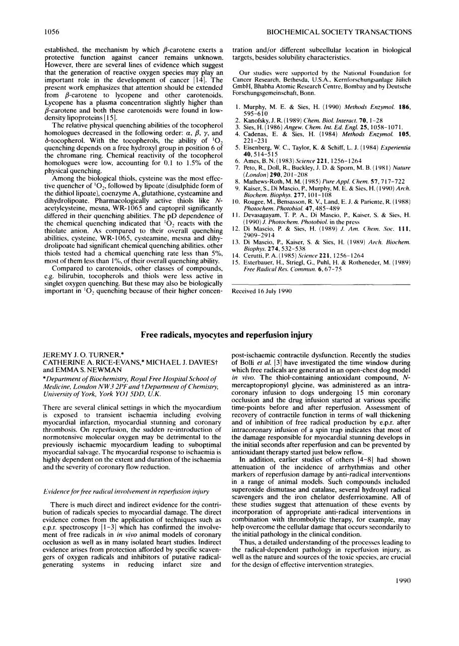
Table 1 . Singlet oxygen yirenching cotistants, chemit.ri1 reaction
rate constants and cotitetit in matntiicrlicin tisstres of ccirotenoidv,
bile pigments, tocopherols, thiols rind related cornpounds
The ( k , + k , ) values were obtained from Stern-Volmcr plots. as
exemplified by Fig. 1 (see Materials and methods). ND - not
determined. From (Y. 11, 131; singlet oxygen lifetime (10 ps)
used for calculations.
B-Carotene
Compound
5
Content i n tissues
'
~
J
-
Lutein
Lycopcnc
y-Carotene
Astaxanthin
Canthaxanthin
a-Carotene
/j-Carotene
Bixin
Zeaxanthin
Lutcin
Cryptoxanthin
Biliruhin
0
0
5
Molarity (
10
p ~ )
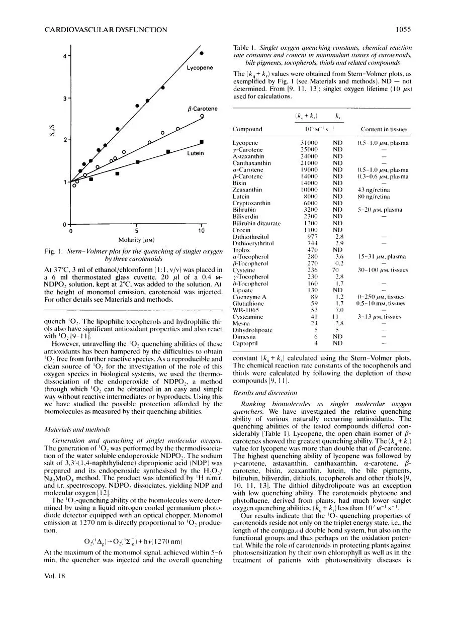
Fig. 1 . Sterti- Volmer plot for the qiierichirig of siriglet oxygen
by three curoterioids
At 37°C. 3 ml of ethanol/chloroform ( 1 : I , V / V ) was placed in
a 6 ml thermostated glass cuvette. 2 0 pI of a 0.4 MNDPO, solution. kept at 2°C. was added to the solution. At
the height of monomol emission, carotenoid was injected.
For other details see Materials and methods.
quench '0,.T h e lipophilic tocopherol5 and hydrophilic thiols also have significant antioxidant properties and also react
with '0,( Y - I 11.
However. unravelling the '0, quenching abilities of thcsc
antioxidants has been hampered by the difficulties t o obtain
'02free from further reactive species. A s a reproducible and
clean source o f '0: for the investigation of the role o f this
oxygen species in biological systems. we used thc thermodissociation o f the endoperoxide o f NDPO,, a method
through which '0, can be obtained in an casy and simple
way without reactive intermediates or byproducts. Using this
we have studied the possible protection afforded by the
biomolccules as measured by their quenching abilities.
Mritcrirr1.s rrtitl methor1.s
(;etierritioti ( i t i d qireti~~hitig
01' siriglct molec~rrlcrr0sj:qcri.
T h e generation o f '0,was performed by the thermodissociation of the water soluble endoperoxide NDPO,. T h e sodium
salt o f 3.3'-( 1.4-naphthylidene) dipropionic acid ( N D P ) was
prepared and its endoperoxide synthcsised by the H,O,/
Na,MoO, method. T h e product was identified by ' H n.m.r.
and i.r. spectroscopy. NDPO, dissociates, yielding N D P and
molecular oxygen [ 131.
T h e '0,-quenching ability of the biomolecules were determined by using a liquid nitrogen-cooled germanium photodiode detector cquippcd with an optical chopper. Monomol
emission at I170 nm is directly proportional to ' 0 , production.
At the maximum of the monomol signal. achievcd within S-6
min. the quenchcr was injcctcd and the overall quenching
VOl. 1 x
Bilivcrdin
Hiliruhin ditaurntc
Crocin
Dithiothreitol
Dit hiocryt hritol
Trolox
a-Tocopherol
/$Tocopherol
Cy5teine
y-Tocop herd
h-l'ocopherol
Lipoatc
Coenzyme A
Glutat hione
w I<- I 005
Cystcaniinc
Mcsna
I>i h yd rolipoatc
Dimesnn
Ciiptopril
3 I000
25000
24000
2 I000
I9000
I4000
I3000
I0000
8000
0000
3200
2300
I200
I I00
977
744
470
2x0
270
236
230
I00
I30
89
59
53
41
24
N I>
N I>
NI)
N I>
N I>
NI)
NI>
N I>
NI>
NI>
NI1
NI)
NI)
NI)
2.8
2.0
NI>
3.0
0 , s - I .O I ~ M .plasma
-
0 . 5 - I .O ,UM, plasma
0.3-0.6 ,UM. plasma
43 ng/rctinn
80 ng/retin;i
5-20
~ I M plasma
,
-
15-3 I
0.2
70
2.8
I .7
NI)
I .2
I .7
7.0
I1
2.8
~ I M plama
,
-
30- I00 ~
I M tissucs
,
-
0-250
I ~ M ,tihsues
0.5- 10 mM. tissues
-
3- I3 / I M . tissues
-
5
i
-
6
NI)
NI>
-
4
-
constant (k,,+ k , ) calculated using the Stern-Volmer plots.
T h e chcmical reaction rate constants o f the tocopherols and
thiols were calculnted by following the depletion o f thcsc
compounds [y, 1 11.
Re.sir1t.s utid di.scri.s.siori
Kmkirig hiornolecirles 11s singlet molectrlur oxygeri
qirericher.~.We have investigated the relative quenching
ability o f various naturally occurring antioxidants. T h e
quenching abilities o f the tested cornpounds differed considerably (Table I ) . Lycopene, the open chain isomer o f /3carotenes showed thc greatest quenching ability. T h e ( k , + k , )
value for lycopene was more than double that o f @-carotene.
T h e highest quenching ability of lycopene was followed by
y-carotene, astaxanthin. canthaxanthin. a-carotene. Bcarotene, bixin, zcaxanthin. lutcin, the bilc pigments,
bilirubin, biliverdin, dithiols, tocopherols and other thiols [Y,
10. I I , 131. T h c dithiol dihydrolipoatc was an cxception
with low quenching ability. T h e carotenoids phytocne and
phytofluene, derived from plants, had much lower singlet
oxygen quenching abilities, (k, + k , ) less than 10' M - I s- I.
quenching properties o f
Our results indicate that the
carotenoids reside not only on the triplet energy state. i.e., the
length o f the conjuga.( d double bond system, but also o n the
functional groups and thus perhaps on the oxidation potential. While the role of carotenoids in protecting plants against
photosensitization by their own chlorophyll as well a s in thc
treatment o f patients with photosensitivity diseases is
1056
BIOCHEMICAL SOCIETY TRANSACTIONS
established, the mechanism by which /?-carotene exerts a
protective function against cancer remains unknown.
However, there are several lines of evidence which suggest
that the generation of reactive oxygen species may play an
important role in the development of cancer [14]. The
present work emphasizes that attention should be extended
from /?-carotene to lycopene and other carotenoids.
Lycopene has a plasma concentration slightly higher than
/?-carotene and both these carotenoids were found in lowdensity lipoproteins [ IS].
The relative physical quenching abilities of the tocopherol
homologues decreased in the following order: a,/?,y, and
&tocopherol. With the tocopherols, the ability of ‘02
quenching depends on a free hydroxyl group in position 6 of
the chromane ring. Chemical reactivity of the tocopherol
homologues were low, accounting for 0.1 to 1.5”/0 of the
physical quenching.
Among the biological thiols, cysteine was the most effective quencher of lo,,followed by lipoate (disulphide form of
the dithiol lipoate), coenzyme A, glutathione, cysteamine and
dihydrolipoate. Pharmacologically active thiols like Nacetylcysteine, mesna, WR- 1065 and captopril significantly
differed in their quenching abilities. The p D dependence of
the chemical quenching indicated that ‘0,reacts with the
thiolate anion. As compared to their overall quenching
abilities, cysteine, WR- 1065, cysteamine, mesna and dihydrolipoate had significant chemical quenching abilities. other
thiols tested had a chemical quenching rate less than S%,
most of them less than I%, of their overall quenching ability.
Compared to carotenoids, other classes of compounds,
e.g. bilirubin, tocopherols and thiols were less active in
singlet oxygen quenching. But these may also be biologically
important in ’02
quenching because of their higher concen-
tration and/or different subcellular location in biological
targets, besides solubility characteristics.
Our studies were supported by the National Foundation for
Cancer Research, Bethesda, U.S.A., Kernf(,rschungsanlage Jiilich
GmbH, Bhabha Atomic Research Centre, Bombay and by Deutsche
Forschungsgemeinschaft, Bonn.
1. Murphy, M. E. & Sies, H. (1990)Methods Enzyrnol. 186,
595-6 10
2. Kanofsky, J. R. (1989) Chern. Biol.Interact. 70, 1-28
3. Sies, H.(1986)Angew. Chern. Int. Ed. Engl. 25, 1058-1071.
4. Cadenas, E. & Sies, H. (1984) Methods Enzymol. 105,
221-231
5 . Eisenberg, W. C., Taylor, K. & Schiff, L. J. ( 1984)Experientiu
40,514-515
1256-1264
6. Ames,B.N.(1983).Scirnce221,
7. Peto, R., Doll, R., Buckley, J. D. & Sporn, M. B.( 198 I ) Nuture
(London)290,201-208
8. Mathews-Roth, M. M. ( 1 9 8 5 ) Pure Appl. C’hern.57,717-722
9. Kaiser, S.,Di Mascio, P.. Murphy, M. E. & Sies. H. ( 1990) Arch.
Biochern. Biophys. 277, I 0 I - 108
10. Rougee, M., Bensasson, R. V., Land, E. J. & Pariente, R. (1988)
Photochem. I’hotobiol. 47,485-489
11. Devasagayam, T. P. A,, Di Mascio, P.. Kaiser, S. & Sies, H.
( 1990)J . I’hotochern. I’hotohiol. in the press
12. Di Mascio, P. & Sies. H. (1989)J. Am. C’hem. Soc. 111,
2909-29I4
13. Di Mascio, P., Kaiser, S. & Sies, H. ( 1 989) Arch. Hiochern.
Biophys. 274,532-538
14. Cerutti, P. A. ( 1985) Science 221, 1256- I264
15. Esterbauer, H., Striegl, G., Puhl, H. & Rotheneder, M. (1989)
Free Hadicul Hex Cbmrnitn. 6.67-75
Received I6 July 1990
Free radicals, myocytes and reperfusion injury
JEREMY J. 0.TURNER,*
CATHERINE A. RICE-EVANS,* MICHAEL J. DAVIESt
and EMMA S. NEWMAN
*Department of Biochemistry, Royal Free Hospital School of
Medicine, London N W.3 217; und ?Department of Chemistry,
University of York, York YO1 5DD, U . K .
There are several clinical settings in which the myocardium
is exposed to transient ischaemia including evolving
myocardial infarction, myocardial stunning and coronary
thrombosis. On reperfusion, the sudden re-introduction of
normotensive molecular oxygen may be detrimental to the
previously ischaemic myocardium leading to suboptimal
myocardial salvage. The myocardial response to ischaemia is
highly dependent on the extent and duration of the ischaemia
and the severity of coronary flow reduction.
Evidence for free rudicul involvement in repetjirsion injirry
There is much direct and indirect evidence for the contribution of radicals species to myocardial damage. The direct
evidence comes from the application of techniques such as
e.p.r. spectroscopy [ 1-31 which has confirmed the involvement of free radicals in in vivo animal models of coronary
occlusion as well as in many isolated heart studies. Indirect
evidence arises from protection afforded by specific scavengers of oxygen radicals and inhibitors of putative radicalgenerating systems in reducing infarct size and
post-ischaemic contractile dysfunction. Recently the studies
of Bolli et al. [3]have investigated the time window during
which free radicals are generated in an open-chest dog model
in vivo. The thiol-containing antioxidant compound, Nmercaptopropionyl glycine, was administered as an intracoronary infusion to dogs undergoing 15 min coronary
occlusion and the drug infusion started at various specific
time-points before and after reperfusion. Assessment of
recovery of contractile function in terms of wall thickening
and of inhibition of free radical production by e.p.r. after
intracoronary infusion of a spin trap indicates that most of
the damage responsible for myocardial stunning develops in
the initial seconds after reperfusion and can be prevented by
antioxidant therapy started just below reflow.
In addition, earlier studies of others [4-81 had shown
attenuation of the incidence of arrhythmias and other
markers of reperfusion damage by anti-radical interventions
in a range of animal models. Such compounds included
superoxide dismutase and catalase, several hydroxyl radical
scavengers and the iron chelator desferrioxamine. All of
these studies suggest that attenuation of these events by
incorporation of appropriate anti-radical interventions in
combination with thrombolytic therapy, for example, may
help overcome the cellular damage that occurs secondarily to
the initial pathology in the clinical condition.
Thus, a detailed understanding of the processes leading to
the radical-dependent pathology in reperfusion injury, as
well as the nature and sources of the toxic species, are crucial
for the design of effective intervention strategies.
1000
Download 1054.full
1054.full.pdf (PDF, 352.46 KB)
Download PDF
Share this file on social networks
Link to this page
Permanent link
Use the permanent link to the download page to share your document on Facebook, Twitter, LinkedIn, or directly with a contact by e-Mail, Messenger, Whatsapp, Line..
Short link
Use the short link to share your document on Twitter or by text message (SMS)
HTML Code
Copy the following HTML code to share your document on a Website or Blog
QR Code to this page

This file has been shared publicly by a user of PDF Archive.
Document ID: 0000594028.