JDIT 2014 1030 006 (PDF)
File information
Title:
Author: Sean
This PDF 1.3 document has been generated by Microsoft® Word 2010 / http://www.convertapi.com, and has been sent on pdf-archive.com on 30/05/2017 at 00:32, from IP address 90.218.x.x.
The current document download page has been viewed 459 times.
File size: 272.08 KB (22 pages).
Privacy: public file


File preview
Journal of Diagnostic Imaging in Therapy. 2014; 1(1): 81-102
Giovannini et al.
Open Medscience
Peer-Reviewed Open Access
JOURNAL OF DIAGNOSTIC IMAGING IN THERAPY
Journal homepage: www.openmedscience.com
Review Article
68
Ga-Somatostatin
Tumors
Analogue
PET/CT
in
Neuroendocrine
Elisabetta Giovannini*, Maria Chiara Gaeta, Andrea Ciarmiello
Nuclear Medicine Unit, Sant’Andrea Hospital, La Spezia, Italy
*Author to whom correspondence should be addressed:
Elisabetta Giovannini, M.D.
elisabetta.giovannini@asl5.liguria.it
Abstract
Neuroendocrine tumors (NETs) include a spectrum of neoplasms characterized by histologic
heterogeneity with significant clinical differences. Generally are well differentiated tumors but often
present metastases at diagnosis. Conventional imaging techniques result insufficient in early diagnosis
and therapy monitoring. Standardized morphological criteria to assess treatment response are
inadequate in NETs, because of their biologic evolution and the cytostatic nature of new oncologic
treatments. Functional imaging modalities have improved the understanding and diagnosis of NETs by
the use of somatostatin analogue tracers labelled with radioisotopes. 111In-Octreotide scintigraphy was
considered the gold standard imaging modalities for NET detection with a diagnostic accuracy
approximately of 90%. Actually 68Ga-Dota-SST radiotracers (SSTRTs) PET/CT represent a superior
imaging procedure with higher accuracy in detection of NET lesions as compared to morphological
imaging procedures and somatostatin receptor scintigraphy. Additionally, the use of somatostatin
analogue radiolabelled tracers offers the possibility to non-invasively evaluate the presence of
somatostatin receptor expression on NET cells, with direct therapeutic implications. However, in the
management of patients with NETs and in the evaluation of response to therapy the specialists
ISSN: 2057-3782 (Online)
http://dx.doi.org/10.17229/jdit.2014-1030-006
81
Journal of Diagnostic Imaging in Therapy. 2014; 1(1): 81-102
Giovannini et al.
opinions remain various. This review is based on a PubMed search of medical literature and presents
the systems of classification, grading and staging of NETs. It also summarizes common
recommendations for the management of patients with NETs, focused especially on the role of 68GaDOTA-SSTRTs PET/CT. In this review, the function of 68Ga-DOTA-SSTRTs PET/CT in pediatric
neuroendocrine tumors is also explored.
Keywords: neuroendocrine tumors, 68Ga-DOTA-SSTRTs PET/CT, somatostatin receptor.
Introduction
Neuroendocrine neoplasms (NETs) are tumors with an incidence of approximately 5/100,000 per year.
Annual incidence is increasing over the past decades due to improvements in diagnosis and the
availability of a more sensitive biochemical evaluation. Therefore, although NETs are traditionally
considered rare, their prevalence is higher than that for gastric, esophageal and pancreatic cancers.
Despite the improved diagnostic workup, making a diagnosis of NETs is usually challenging and often
delayed. As a result metastatic disease at diagnosis is common. The detection of NETs by conventional
imaging, such as computed tomography (CT), ultrasonography (US), and magnetic resonance imaging
(MRI) is limited [1]. Moreover, even in those patients with localized disease, the prognosis is not
favourable with a five-year survival rate of less than 80% because of a high risk of recurrence in a not
negligible proportion of patients [2-3].
Biology
Neuroendocrine neoplasms originate from neuroectoderma or endoderma cells and have both neural
and endocrine cell features. It is well known that NETs can develop in almost all tissues or organs in
the body, especially in the gastroenteropancreatic tract and lungs and rarely in the ovary [2]. They are
characterized by an ample range of histological appearance and biological behavior These tumors
show tissue immunoreactivity for markers of neuroendocrine differentiation (synaptophysin,
chromogranin A, CD56, and NSE) and may secrete various peptides and hormones [4]. NETs are
frequently placed in the submucosa or more deeply intramurally and can invade surrounding tissues.
The tumoral cells can be organized variously in islands, glands or sheets.
Often neuroendocrine cells show minimal pleomorphism but they can evolve in anaplasia andrapid
mitotic activity leading to intratumoral areas of necrosis. Some neuroendocrine tumor cells express
hormone receptors with a strong avidity for specific hormones that can be exploited for diagnosis and
treatment purposes. In particular, neuroendocrine tumors possess somatostatin receptors (SSTRs) with
a very variable expression in terms of density and subtypes. Five subtypes of SSTRs, SSTRs 1-5; have
been cloned and they belong to a distinct group within the superfamily of G-protein-coupled receptors
with seven transmembrane regions [5].
A high density of somatostatin receptors is found in neuroendocrine tumors, such as pituitary
adenoma, gastroentropancreatic tumors, carcinoid, pheochromocytoma, paraganglioma, medullary
ISSN: 2057-3782 (Online)
http://dx.doi.org/10.17229/jdit.2014-1030-006
82
Journal of Diagnostic Imaging in Therapy. 2014; 1(1): 81-102
Giovannini et al.
thyroid cancer, and small cell lung carcinoma. Tumors of the nervous system including meningioma,
neuroblastoma, and medulloblastoma also often express a high density of SSTR. Tumors not
originating from the neural or endocrine cells, such as lymphoma, breast cancer, renal cell cancer,
hepatocellular carcinoma, prostate cancer, sarcoma and gastric cancer, may also express SSTR. The
expression of SSTR is not specific for malignancy. Some benign lesions may also express SSTR, for
example, active granulomas in sarcoidosis [6].
Classification of Neuroendocrine Tumors
A consensus about a recognized uniform grading system for neuroendocrine neoplasms has been
difficult to achieve. The systems proffered by the American Joint Committee on Cancer (AJCC),
World Health Organization (WHO), European Neuroendocrine Tumor Society (ENETS) and others
provide useful prognostic information [1, 7].
Neuroendocrine neoplasms can be classified by: anatomic site of origin; histology: well-differentiated
(G1 and G2) NETs, poorly differentiated (G3) carcinomas or undifferentiated neoplasms; extent of
disease: local, regional or distant metastases.
Most NETs arise from the GI tract (stomach, appendix, duodenum, and small intestine), the
bronchopulmonary system (lungs and thymus), the pancreas, the colon and rectum. The biological
behaviour exhibited by neuroendocrine neoplasms is highly correlated with neoplasm grade. The grade
of a tumor refers to its biologic aggressiveness. The grading system is based on the rate of
proliferation, which is defined by the number of mitoses per 10 high-power microscopic fields or per 2
mm2 (mitotic rate), or as the percentage of tumor cells that immunolabel positively for the Ki-67
antigen (Ki-67 index). Briefly, low-grade tumors are characterized by low proliferative indices and are
considered indolent in nature. High-grade tumors tend to be poorly differentiated, have high
proliferative indices and are therefore very aggressive [8] (Table1).
Grade
Tumor
Lung, Thymus (WHO)
GEP-NETs (ENETS, WHO)
Low
<2 mitoses/10 hpf, AND no necrosis
<2 mitoses/10 hpf, AND Ki67 index <3%
Intermediate
2–10 mitoses/10 hpf OR foci of necrosis
2–20 mitoses/10 hpf, OR Ki67 index 3%–20%
High
>10 mitoses/10 hpf
>20 mitoses/10 hpf OR Ki67 index >20%
Abbreviations: WHO (World Health Organization); GEP (gastroenteropancreatic); NETs (neuroendocrine tumors);
ENETS (European Neuroendocrine Tumor Society); hpf (high-power fields).
Table 1. Grading Systems for Neuroendocrine.
ISSN: 2057-3782 (Online)
http://dx.doi.org/10.17229/jdit.2014-1030-006
83
Journal of Diagnostic Imaging in Therapy. 2014; 1(1): 81-102
Giovannini et al.
For neuroendocrine neoplasms, the presence of necrosis also plays an important role in grading. For
example, G1/G2 bronchial NETs (typical/atypical carcinoids) and G3 bronchial neoplasms (large cell
neuroendocrine carcinoma [LCNEC] and small cell lung carcinoma [SCLC]) exhibit markedly
different behavior. The presence of necrosis, alongside mitotic activity, is a distinguishing feature
between these 2 groups of tumors. NETs can also be classified based on differentiation, referring to the
extent to which neoplastic cells resemble normal cells.
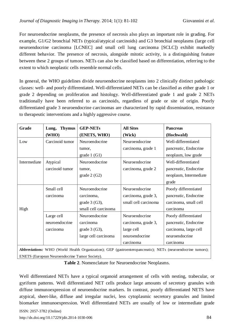
In general, the WHO guidelines divide neuroendocrine neoplasms into 2 clinically distinct pathologic
classes: well- and poorly differentiated. Well-differentiated NETs can be classified as either grade 1 or
grade 2 depending on proliferation and histology. Well-differentiated grade 1 and grade 2 NETs
traditionally have been referred to as carcinoids, regardless of grade or site of origin. Poorly
differentiated grade 3 neuroendocrine carcinomas are characterized by rapid dissemination, resistance
to therapeutic interventions and a highly aggressive course.
Grade
Low
Lung, Thymus GEP-NETs
All Sites
Pancreas
(WHO)
(ENETS, WHO)
(Wick)
(Hochwald)
Carcinoid tumor
Neuroendocrine
Neuroendocrine
Well-differentiated
tumor,
carcinoma, grade 1
pancreatic, Endocrine
grade 1 (G1)
Intermediate Atypical
carcinoid tumor
neoplasm, low grade
Neuroendocrine
Neuroendocrine
Well-differentiated
tumor,
carcinoma, grade 2
pancreatic, Endocrine
grade 2 (G2)
neoplasm, Intermediate
grade
Small cell
Neuroendocrine
Neuroendocrine
Poorly differentiated
carcinoma
carcinoma,
carcinoma, grade 3,
pancreatic, Endocrine
grade 3 (G3),
small cell carcinoma
carcinoma, small cell
High
small cell carcinoma
carcinoma
Large cell
Neuroendocrine
Neuroendocrine
Poorly differentiated
neuroendocrine
carcinoma
carcinoma, grade 3,
pancreatic, Endocrine
carcinoma
grade 3 (G3),
large cell
carcinoma, large cell
large cell carcinoma
neuroendocrine
neuroendocrine
carcinoma
carcinoma
Abbreviations: WHO (World Health Organization); GEP (gastroenteropancreatic); NETs (neuroendocrine tumors);
ENETS (European Neuroendocrine Tumor Society).
Table 2. Nomenclature for Neuroendocrine Neoplasms.
Well differentiated NETs have a typical organoid arrangement of cells with nesting, trabecular, or
gyriform patterns. Well differentiated NET cells produce large amounts of secretory granules with
diffuse immunoexpression of neuroendocrine markers. In contrast, poorly differentiated NETS have
atypical, sheet-like, diffuse and irregular nuclei, less cytoplasmic secretory granules and limited
biomarker immunoexpression. Well differentiated NETs are usually of low or intermediate grade
ISSN: 2057-3782 (Online)
http://dx.doi.org/10.17229/jdit.2014-1030-006
84
Journal of Diagnostic Imaging in Therapy. 2014; 1(1): 81-102
Giovannini et al.
whereas poorly differentiated NETs are usually high grade. The histological classification of NETs,
including grade (G) and differentiation is outlined in Table 2.
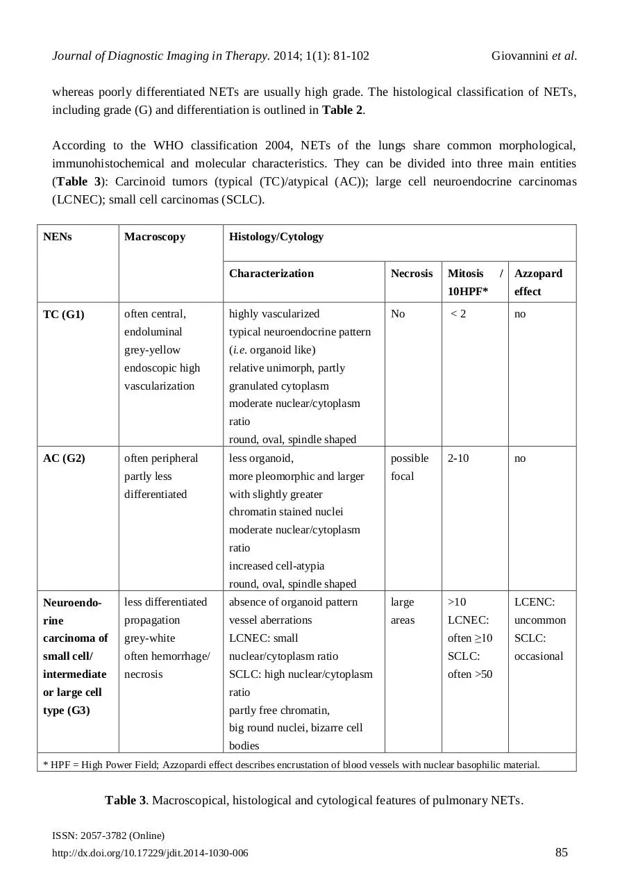
According to the WHO classification 2004, NETs of the lungs share common morphological,
immunohistochemical and molecular characteristics. They can be divided into three main entities
(Table 3): Carcinoid tumors (typical (TC)/atypical (AC)); large cell neuroendocrine carcinomas
(LCNEC); small cell carcinomas (SCLC).
NENs
Macroscopy
Histology/Cytology
Characterization
TC (G1)
often central,
highly vascularized
endoluminal
typical neuroendocrine pattern
grey-yellow
(i.e. organoid like)
endoscopic high
relative unimorph, partly
vascularization
granulated cytoplasm
Necrosis
Mitosis
/ Azzopard
10HPF*
effect
No
<2
no
2-10
no
moderate nuclear/cytoplasm
ratio
round, oval, spindle shaped
AC (G2)
often peripheral
less organoid,
possible
partly less
more pleomorphic and larger
focal
differentiated
with slightly greater
chromatin stained nuclei
moderate nuclear/cytoplasm
ratio
increased cell-atypia
round, oval, spindle shaped
Neuroendo-
less differentiated
absence of organoid pattern
large
>10
LCENC:
rine
propagation
vessel aberrations
areas
LCNEC:
uncommon
carcinoma of
grey-white
LCNEC: small
often ≥10
SCLC:
small cell/
often hemorrhage/
nuclear/cytoplasm ratio
SCLC:
occasional
intermediate
necrosis
SCLC: high nuclear/cytoplasm
often >50
or large cell
ratio
type (G3)
partly free chromatin,
big round nuclei, bizarre cell
bodies
* HPF = High Power Field; Azzopardi effect describes encrustation of blood vessels with nuclear basophilic material.
Table 3. Macroscopical, histological and cytological features of pulmonary NETs.
ISSN: 2057-3782 (Online)
http://dx.doi.org/10.17229/jdit.2014-1030-006
85
Journal of Diagnostic Imaging in Therapy. 2014; 1(1): 81-102
Giovannini et al.
These neuroendocrine entities are further classified into two groups; according to their biological
aggressiveness [9]: well-differentiated low grade (G1) typical and intermediate grade (G2); atypical
carcinoids; poorly-differentiated high grade (G3) LCNEC and SCLC. In contrast with typical and
atypical carcinoids, LCNEC and SCLC are not closely related to each other regarding the genetic and
epigenetic characteristics. Unlike carcinoids, no precursor lesions are known for SCLCs and LCNECs
[10-12].
In typical carcinoids, regional lymph node metastases can be found in 10-15% and distant metastases
in 3-5% [13]. In atypical carcinoids, nodal metastases are found in 50% and distant metastases in 25%.
As metastases of G1 and G2 NETs are not sensitive to chemotherapy, surgery remains the first choice
of treatment in metastatic diseases. Due to the high metastatic risk (in 50% to 80%) of these tumors,
prophylactic cranial radiation is usually done [14].
Small cell lung cancers because of differences in clinical behavior, therapy and epidemiology are
classified separately. Several staging systems have been proposed for small cell lung cancer (SCLC)
by American Joint Committee on Cancer (AJCC), WHO, Veterans Administration Lung Study Group
(VALG) and International Association for the Study of Lung Cancer (IASLC) [15-16]. The staging
systems include two levels according to metastasis: Limited-Stage Disease (LD) (disease is confined to
the hemithorax of origin, the mediastinum or the supraclavicular nodes, which can be encompassed
within a tolerable radiation therapy port).
Patients with pleural effusion, massive pulmonary tumor, and contralateral supraclavicular nodes have
been both included within and excluded from LD by various groups. Extensive-Stage Disease (ED)
(SCLC has spread beyond the supraclavicular areas and is too widespread to be included within the
definition of LD. Patients with distant metastases (M1) are always considered to have ED) [15-16].
This distinction is crucial because the shape of limited disease responds very well to chemotherapy
associated with radiotherapy, especially with drug combinations that use the cis-platinum. In general
surgery is not recommended [17].
More recently the WHO classification of the neuroendocrine neoplasms of gastroentropancreatic
(GEP) system subdivided them into two major categories: neuroendocrine tumor (NET) and
neuroendocrine carcinoma (NEC), based on histological differentiation (well differentiated and poorly
differentiated), proliferative activity (G1, G2 and G3) and TNM factors (size, infiltration/invasion and
metastasis). NET G1 and NET G2 correspond to what were formerly called well differentiated
endocrine tumor and well differentiated endocrine carcinoma, respectively. NEC G3 is nearly the same
as poorly differentiated endocrine carcinoma and highly malignant [18-19] Table 2.
The European Neuroendocrine Tumor Society and the American Joint Committee on Cancer stage
NETs by the primary tumor (T), lymph node involvement (N), and distant metastasis (M). This TNM
staging represents a new system of NET classification. The definition of TNM varies by each primary
tumor site; however, staging relies predominantly on the size of the tumor and the extent of invasion
ISSN: 2057-3782 (Online)
http://dx.doi.org/10.17229/jdit.2014-1030-006
86
Journal of Diagnostic Imaging in Therapy. 2014; 1(1): 81-102
Giovannini et al.
into anatomical structures. For example, a NET in the colon invading the muscularis propria with no
lymph node involvement or distant metastases would be considered Stage IIA or IIB.
The North American Neuroendocrine Tumor Society (NANETS) recommend that pathology reports
provide a TNM stage, based on a system that is specifically referenced in the pathology report [19-20].
In the NET G1, G2 group, SSTR-2A expression is significantly higher in the gastrointestinal primaries
than in the lung primaries. This may suggest that SSTR-2A is a receptor subtype, characterizing the
low grade neuroendocrine neoplasms of GI origin. Among the SSTR subtypes, the expression of
SSTR-2A was significantly high in both the NET and NEC groups. No significant difference was
observed in the expression pattern of SSTR subtypes in the NET and NEC groups.
However, a significant difference in the expression profiles of SSTR-1 and 2A between NEC G3 small
cell type and non-small cell type was obtained. In NEC G3 small cell type, the expression of SSTR-1
and 2A was significantly low. Alternatively, the expression of SSTR-5 is rather high leading to the
conclusion that NEC G3 should be classified into small cell type and non-small cell type for example
lung cancer.
All the specific antibodies against SSTR-1, 2A, 3 and 5 show specific and satisfactory
immunolocalization of SSTR subtypes in the tumor cells. The immunolocalization of SSTR-2A is
usually membranous and intensely stained. Regarding the expression of SSTR-2A in the NET G1, G2
group, there is relatively good correlation between the expressions of mRNA (100%) and protein
(>80%). However, in the NEC G3 group the expression of SSTR-2A protein is rather low (61.9%)
compared with the expression of mRNA (95.2%). Such discrepant results may be caused by the
different sensitivities of each detection system.
The immunohistochemical localization of SSTR-1, 3 and 5 is usually cytoplasmic, but membranous
localization is also seen occasionally. No significant correlation is observed between the expression of
neuroendocrine markers (synaptophysin, chromogranin A, CD56, and NSE) and the expression
patterns of SSTR subtypes [21]. The expression of SSTR represents the bases of molecular imaging.
Imaging Modalities
Morphological or structural imaging technologies available for assessment of NETs include CT, MRI,
and ultrasonography. These imaging techniques are used especially to assess the morphologic features,
to identify changes in the target lesions over time and to detect new tumors through use of serial
imaging. Each of these modalities has advantages and disadvantages dependent by tumor characteristic
and location.
Computed Tomography
Computed tomography (CT) is the modality of choice for the initial imaging work-up and for therapy
monitoring of NET disease. Radiologically, pulmonary carcinoids (TC ≤ 2 cm and AC ≥ 4 cm) are
nodules or masses (TC with a smooth margin and AC with an irregular margin). Approximately 30ISSN: 2057-3782 (Online)
http://dx.doi.org/10.17229/jdit.2014-1030-006
87
Journal of Diagnostic Imaging in Therapy. 2014; 1(1): 81-102
Giovannini et al.
55% of carcinoids cause lobar atelectasis, obstructive pneumonitis and partial obstruction. Their
visualization can improve with intravenous administration of contrast due to the marked enhancement
of their vascular stroma [22].
In SCLC usually a central mass formed by the combination of primary tumor and lymph node
metastases can be found radiologically. Mediastinal lymph node involvement is present in most cases
(5% to 10% present as a peripheral nodule without lymph node involvement). Reduction and
displacement of major vessels and bronchi and pleural effusion are common findings.
Contrast-enhancement CT examination is especially important in early disease for pre-operative
planning. Three-dimensional (3D) volume rendering technique (VRT) and maximum intensity
projections (MIPs) in CT angiography is useful preoperatively to visualize arterial anatomy and detect
vascular encasement.
The high spatial resolution of current CT scanners allows to show the relation between the pancreatic
duct and tumor in the GEP [23-24]. In general for abdominal imaging, MRI is superior to CT because
of the better soft tissue contrast. On the other hand, the better spatial resolution of CT is more
advantageous for detection of small lung metastases and therefore for examination of the thorax CT
rather than MRI should be performed [25].
Magnetic Resonance Imaging
For MRI, specific imaging protocols for NETs have been described, including hyperintense signal
intensity on fluid-fluid levels on T2-weighted (T2w) imaging and arterial hypervascularity. Intra
vascular contrast enhancement is mandatory in MRI for NETs imaging and it is recommended that the
liver and pancreas should be always examined by so called ‘triple-phase scanning’. The examination is
performed before and during i.v. contrast enhancement in the late arterial phase (portal-venous inflow
phase) and venous (portal-venous) phase. Magnetic resonance cholangiopancreatography (MRCP) is
too generally included in NET evaluation, allowing better visualization of the main bile duct and the
pancreatic duct also respect to CT [26].
Recent studies suggest diffusion-weighted (DW) MR sequences to be very sensitive for the detection
of NET and for characterization of metastases, particularly helping in identify small lesions [27-28].
Additionally, functional MRI examinations (dynamic enhancement contrast MRI: DCE-MRI) with the
introduction of a particular contrast enhancement, gadolinium-EOB-DTPA, has increased sensitivity
and specificity, particularly for the detection of liver metastases. DCE-MRI is able to quantify the
microcirculatory status of the liver prospecting the possibility to go beyond the mere morphologic
assessment [28].
Somatostatin Receptor Imaging
Over-expression of somatostatin receptors (SSTR) is the rationale for molecular imaging in NET. The
receptors are expressed in about 80–90% of NETs [20], particularly in well-differentiated
ISSN: 2057-3782 (Online)
http://dx.doi.org/10.17229/jdit.2014-1030-006
88
Journal of Diagnostic Imaging in Therapy. 2014; 1(1): 81-102
Giovannini et al.
neuroendocrine tumors. All the subtypes of SSTR expressed by NET have affinity for the native
peptide. Hence, the sensitivity of somatostatin receptor studies depends on the density of the SSTR in
the tumor and the type of analogue used. Somatostatin receptors are expressed by many
neuroendocrine and non neuroendocrine cells of the body, so different organs may be imaged by
somatostatin receptor scintigraphy including the liver, spleen, pituitary, thyroid, kidneys, adrenal
glands, salivary glands, stomach wall, bowel [29-30].
Scintigraphy
Scintigraphy (SRS) using 111In-DTPA-octreotide (Octreoscan) is the standard method. Whole-body
images (anterior and posterior views) are acquired at 4 and 24 h and single photon computed emission
tomography (SPECT or SPECT/CT) of the abdomen and thorax is performed 24 h after injection. The
sensitivity and specificity of SRS is varying in different reports, although a review comprising 35
centers and 1200 patients showed median 89% (range 67 -- 100%) detection rate and median 84%
(range 57 -- 93%) sensitivity, but the sensitivity at SRS varies with the tumor type and its anatomical
localization [31-32].
PET/CT-68Ga-DOTA-SSTRTs
The introduction of 68Ga-DOTA-SSTRTs PET/CT for the evaluation of NETs has significantly
improved the diagnostic work-up, previously based only on conventional imaging (CI) modalities
(ultrasound, CT, endoscopy, MRI) and somatostatin receptor scintigraphy (SRS) [33-34]. 68Ga-DOTAconjugate peptides are rapidly cleared from the blood. Maximal tumour activity accumulation is
reached 70+/-20 min post injection. Excretion is almost entirely through the kidneys [35]. Exist
various 68Ga-peptide preparations (68Ga-DOTATOC, 68Ga -DOTANOC, 68Ga DOTATATE) which
show different affinity to the somatostatin receptor subtypes but appear to be similarly effective in
visualizing NETs in patients [6]. 68Ga-DOTATOC binds to SSTR5 with intermediate affinity and
68
Ga-DOTANOC has high affinity to SSTR2, SSTR3, and SSTR5 [36].
Functional imaging by PET/CT with 68Ga-labeled octreotide and octreotate (68Ga-DOTATOC, 68GaDOTATATE, 68Ga-DOTANOC), has shown excellent results in NET patients. In a large number of
studies, PET and PET/CT with these tracers have shown superior to morphologic imaging, mostly in
comparison with CT. A large prospective study also demonstrates a higher accuracy of 68GaDOTATOC in comparison to the anatomical imaging modality, CT, and conventional SRS [37]. The
advantages of PET over SRS are a higher tissue contrast and a better spatial resolution of about 0.5 cm
compared to 1-1.5 cm with SPECT and planar scintigraphy.
Furthermore, with the 68Ga-preparations there are logistical advantages because of their more favorable
kinetics that allows PET imaging already 1 h after injection [2, 38]. The first clinical investigation on
SSTR targeting using 68Ga-DOTATOC PET was published in 2001 by Henze et al. [39], who studied
patients with meningiomas. The hypothesis of the study was that PET imaging with 68Ga-DOTATOC
could help differentiating between meningiomas, neurofibromatosis and metastases, since
meningiomas highly express SSTR2. They imaged three patients with 68Ga-DOTATOC PET, who had
ISSN: 2057-3782 (Online)
http://dx.doi.org/10.17229/jdit.2014-1030-006
89
Download JDIT-2014-1030-006
JDIT-2014-1030-006.pdf (PDF, 272.08 KB)
Download PDF
Share this file on social networks
Link to this page
Permanent link
Use the permanent link to the download page to share your document on Facebook, Twitter, LinkedIn, or directly with a contact by e-Mail, Messenger, Whatsapp, Line..
Short link
Use the short link to share your document on Twitter or by text message (SMS)
HTML Code
Copy the following HTML code to share your document on a Website or Blog
QR Code to this page

This file has been shared publicly by a user of PDF Archive.
Document ID: 0000603696.