Review topics Quiz 1 Bio 110 STUDY GUIDE complete 2 (PDF)
File information
Author: User
This PDF 1.5 document has been generated by Microsoft® Office Word 2007, and has been sent on pdf-archive.com on 15/04/2018 at 02:02, from IP address 99.47.x.x.
The current document download page has been viewed 534 times.
File size: 1.66 MB (12 pages).
Privacy: public file


File preview
Review topics Quiz 1 Bio 110 Human anatomy.
Define:
Anatomy – The study of internal and external structures and relationship between body parts.
Regional Anatomy – Refers to all structures in a specific area of the body, such as head neck or trunk
including the superficial and deep.
Surface Anatomy – Refers to the superficial anatomical markings.
Histology – The study of tissues.
Physiology – The study of how the body functions and its mechanisms.
Embryology - The study of early developmental stages.
Organelle – The anatomical structures of the cell.
Metabolism – All the chemical reactions in the body.
Phospholipid Bilayer – Two layers of phospholipids that are arranged so that the heads are at the
surface and the tails are on the inside. It helps compose the plasmalemma.
Plasmalemma – The cell membrane or outer boundary of the cell.
Stratified Epithelium - Two or more layers of cells above the basal lamina.
Cephalic – Toward the head.
Dorsal – The back or behind.
Langerhans Cells – Wandering macrophages found in the epidermis.
Cells – The smallest living unit in the body.
Cyanosis - A medical condition in which the lips and fingertips of an individual turn blue due to lack of
adequate delivery of oxygen to tissues.
Endocrine Glands – Ductless glands that release hormone secretions directly into the interstitial
fluids, lymph, or blood.
Exocrine Glands – May be either unicellular or multicellular, secretes mucins, enzymes, water, and
waste through ducts to the epithelial surface.
Hyaline Cartilage – Provides stiff but somewhat flexible support and reduces friction between bony
surfaces. Found in between the ribs and in bones of sternum.
Caudal – Toward the tail. (coccyx in humans)
Proximal – Toward an attached base.
Distal – Away from an attached base.
Gluteal Region – The gluteus or buttock.
Cubital Fossa – “Elbow pit” The area on the anterior of the elbow.
Supine – Lying down (face up) in the anatomical position.
Prone – Lying down (face down) in the anatomical position.
Lateral – Away from the midline.
Medial – Toward the midline.
Neuroglial – The supporting cells of the neural tissue. Help protect the neurons and composes 90-95%
of the neural tissue.
Endocytosis – When the cell membrane opens/engulfs taking in large protein/lipid molecules. If the
molecule is solid, the process it called phagocytosis. If the molecule is liquid, the process is called
pinocytosis.
Osmosis – The movement of water from an area of low solute concentration to an area of high solute
concentration.
What are the levels of organization in anatomy?
Chemical/Molecular → Cell → Tissue → Organ → Organ System → Organism
Blood cells are produced in the body primarily at what location?
Bone marrow (red marrow)
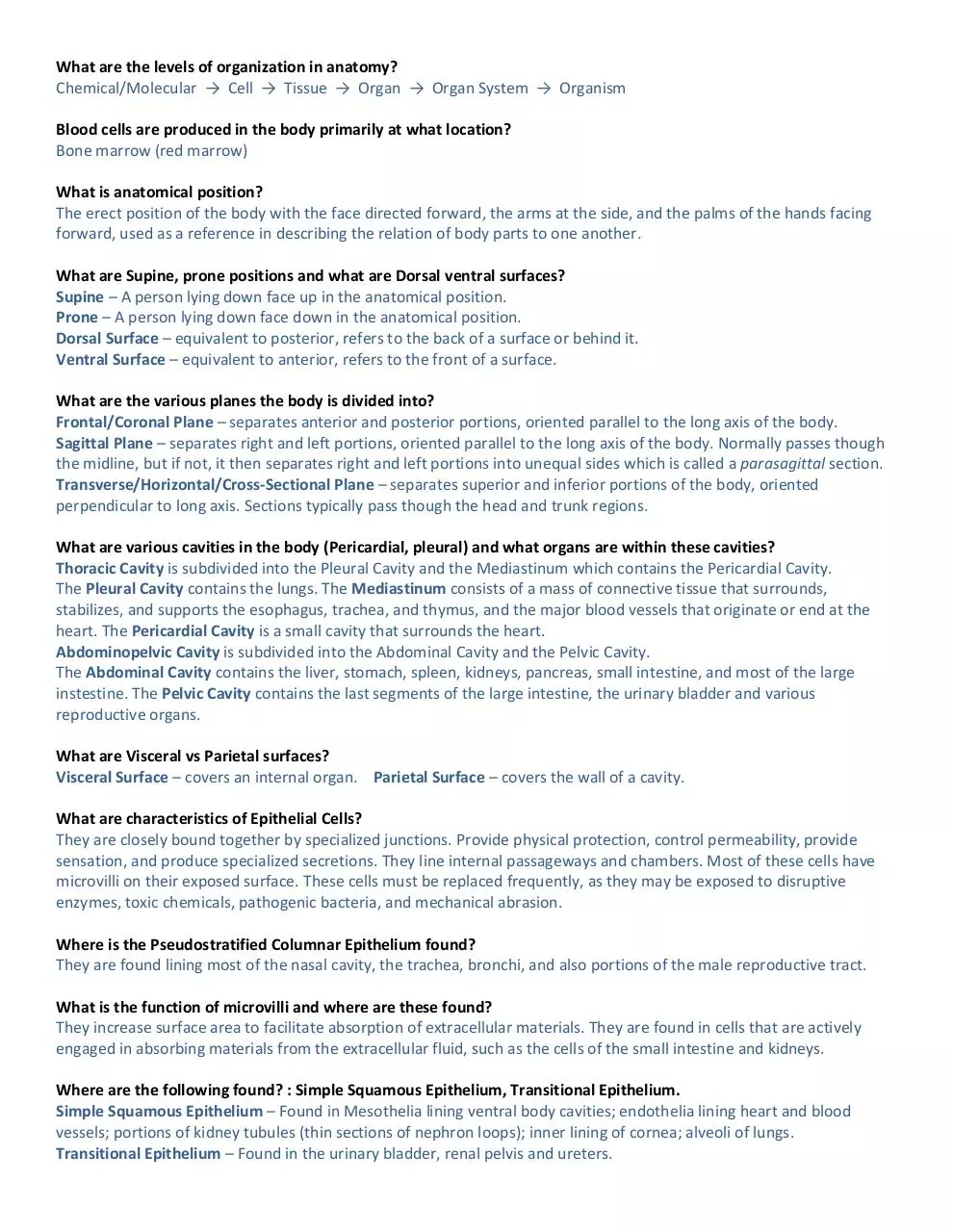
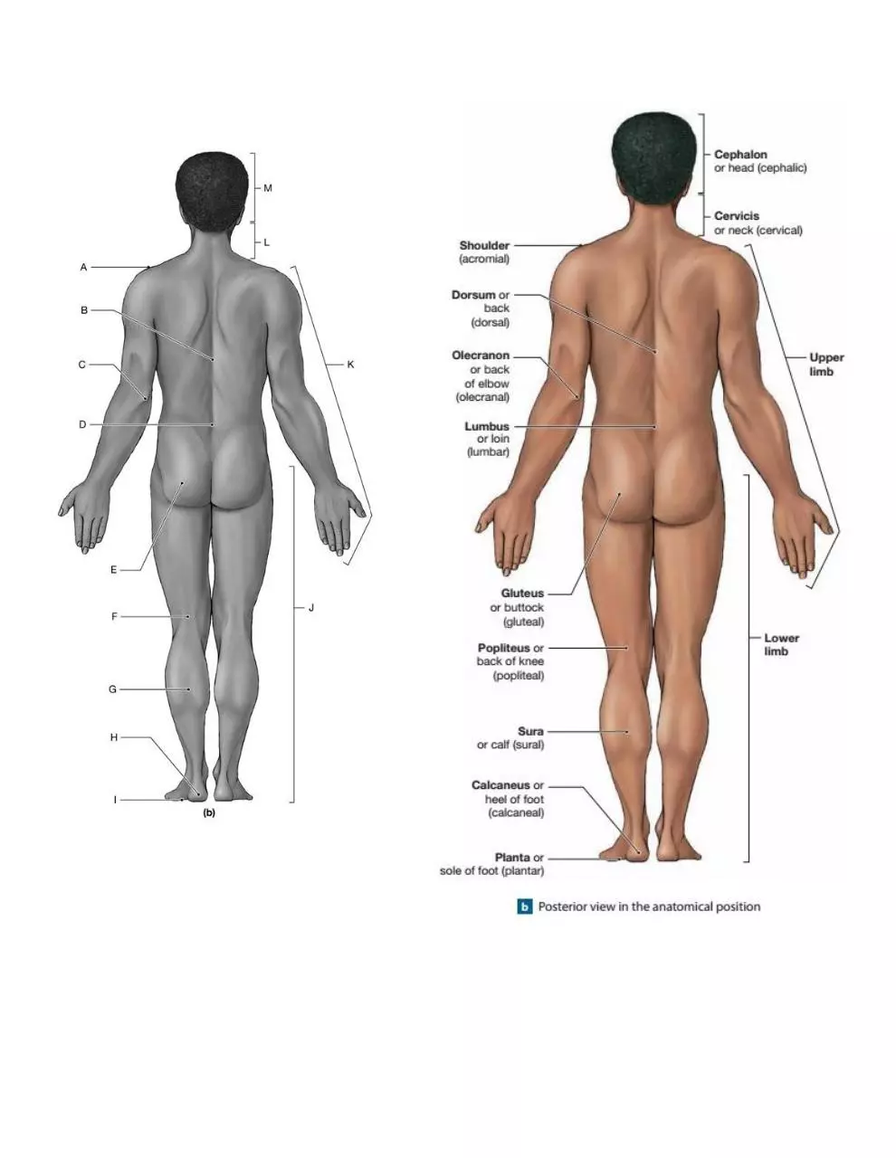
What is anatomical position?
The erect position of the body with the face directed forward, the arms at the side, and the palms of the hands facing
forward, used as a reference in describing the relation of body parts to one another.
What are Supine, prone positions and what are Dorsal ventral surfaces?
Supine – A person lying down face up in the anatomical position.
Prone – A person lying down face down in the anatomical position.
Dorsal Surface – equivalent to posterior, refers to the back of a surface or behind it.
Ventral Surface – equivalent to anterior, refers to the front of a surface.
What are the various planes the body is divided into?
Frontal/Coronal Plane – separates anterior and posterior portions, oriented parallel to the long axis of the body.
Sagittal Plane – separates right and left portions, oriented parallel to the long axis of the body. Normally passes though
the midline, but if not, it then separates right and left portions into unequal sides which is called a parasagittal section.
Transverse/Horizontal/Cross-Sectional Plane – separates superior and inferior portions of the body, oriented
perpendicular to long axis. Sections typically pass though the head and trunk regions.
What are various cavities in the body (Pericardial, pleural) and what organs are within these cavities?
Thoracic Cavity is subdivided into the Pleural Cavity and the Mediastinum which contains the Pericardial Cavity.
The Pleural Cavity contains the lungs. The Mediastinum consists of a mass of connective tissue that surrounds,
stabilizes, and supports the esophagus, trachea, and thymus, and the major blood vessels that originate or end at the
heart. The Pericardial Cavity is a small cavity that surrounds the heart.
Abdominopelvic Cavity is subdivided into the Abdominal Cavity and the Pelvic Cavity.
The Abdominal Cavity contains the liver, stomach, spleen, kidneys, pancreas, small intestine, and most of the large
instestine. The Pelvic Cavity contains the last segments of the large intestine, the urinary bladder and various
reproductive organs.
What are Visceral vs Parietal surfaces?
Visceral Surface – covers an internal organ. Parietal Surface – covers the wall of a cavity.
What are characteristics of Epithelial Cells?
They are closely bound together by specialized junctions. Provide physical protection, control permeability, provide
sensation, and produce specialized secretions. They line internal passageways and chambers. Most of these cells have
microvilli on their exposed surface. These cells must be replaced frequently, as they may be exposed to disruptive
enzymes, toxic chemicals, pathogenic bacteria, and mechanical abrasion.
Where is the Pseudostratified Columnar Epithelium found?
They are found lining most of the nasal cavity, the trachea, bronchi, and also portions of the male reproductive tract.
What is the function of microvilli and where are these found?
They increase surface area to facilitate absorption of extracellular materials. They are found in cells that are actively
engaged in absorbing materials from the extracellular fluid, such as the cells of the small intestine and kidneys.
Where are the following found? : Simple Squamous Epithelium, Transitional Epithelium.
Simple Squamous Epithelium – Found in Mesothelia lining ventral body cavities; endothelia lining heart and blood
vessels; portions of kidney tubules (thin sections of nephron loops); inner lining of cornea; alveoli of lungs.
Transitional Epithelium – Found in the urinary bladder, renal pelvis and ureters.
What are the names and order of five layers of epidermis?
From top to bottom:
Stratum Corneum → Stratum Lucidum → Stratum Granulosum → Stratum Spinosum → Stratum Basale
What factors contribute to the skin color?
The color of the epidermis is due to a combination of the dermal blood supply, the thickness of the stratum corneum,
and variable quantities of two pigments: carotene and melanin.
What are the two types of sweat glands?
Apocrine Glands – Sweat glands that release their secretions into hair follicles in the axillae (armpits), around the
nipples (areolae), and in the groin area.
Merocrine Glands – Coiled tubular glands that discharge their secretions directly onto the surface of the skin.
Where are sebaceous glands found?
Sebaceous Glands – Normally found in hair follicles. They produce an oily lipid (sebum) that coats hair shafts and the
epidermis.
Sebaceous Follicles – Sebaceous glands that communicate directly with the epidermis and never produce hair. Found on
the skin covering the face, back, chest, nipples, and male sex organs.
What kind of gland produces cerumen what is the function of cerumen?
Produced by the Ceruminous Gland. Cerumen (Ear Wax) together with tiny hairs along the ear canal, helps trap foreign
particles or small insects and keeps them from reaching the eardrum.
What are Merocrine, Holocrine, Apocrine glands give example of each?
Apocrine Glands – Sweat glands that release their secretions into hair follicles in the axillae (armpits), around the
nipples (areolae), and in the groin area.
Merocrine Glands – Coiled tubular glands that discharge their secretions directly onto the surface of the skin. Widely
distributed with palms and soles having the highest numbers.
Holocrine – A type of secretion found in Sabaceous Glands where the secretion destroys the gland cell by bursting.
What are the causes of skin wrinkles?
Age, hormones, and the destructive effects of ultraviolet radiation reduce the thickness and flexibility of the dermis,
producing wrinkles and sagging skin.
What is Parietal and Visceral layer of Serous Membranes?
The subdivisions of the ventral body cavity are lined by Serous Membranes. Firmly attached to the body wall or organs
they cover. The Parietal and Visceral portions of a Serous Membrane are in close contact at all times. Minimizing
friction between these opposing surfaces is the primary function of Serous Membranes.
What type of epithelium is found in the blood vessels?
The Simple Squamous Epithelium lining the heart and all blood vessels is called an Endothelium.
Where is cuboidal epithelium found?
Simple Cuboidal Epithelium – Found in glands, ducts, portions of kidney tubules, and the thyroid gland.
Stratified Cuboidal Epithelium – Quite rare, they are found lining the ducts of sweat glands and in the larger ducts of
some other exocrine glands, such as the mammary glands.
Download Review topics Quiz 1 Bio 110 STUDY GUIDE complete 2
Review topics Quiz 1 Bio 110 STUDY GUIDE complete 2.pdf (PDF, 1.66 MB)
Download PDF
Share this file on social networks
Link to this page
Permanent link
Use the permanent link to the download page to share your document on Facebook, Twitter, LinkedIn, or directly with a contact by e-Mail, Messenger, Whatsapp, Line..
Short link
Use the short link to share your document on Twitter or by text message (SMS)
HTML Code
Copy the following HTML code to share your document on a Website or Blog
QR Code to this page

This file has been shared publicly by a user of PDF Archive.
Document ID: 0000756800.