#1 Surgical Management of Congenital Heart Disease (PDF)
File information
Author: LG
This PDF 1.5 document has been generated by Microsoft® Word 2010 Trial, and has been sent on pdf-archive.com on 14/11/2013 at 15:51, from IP address 180.194.x.x.
The current document download page has been viewed 845 times.
File size: 568.93 KB (7 pages).
Privacy: public file


File preview
Surgical Management of Congenital
Heart Disease – Part 1
November 13, 2013
Dr. Purisima Atas
Group 4
Additional information from Batch Jax’s notes were not included
here (if may ara man gid, gamay lang). You are encouraged to read
their novel-like notes from last year if you wish to.
SAVE SPACE SAVE PAPER SAVE TREES SAVE THE EARTH
General Principles of Treatment
•
•
•
•
•
•
•
•
•
•
•
Most patients who have mild congenital heart
disease require no treatment
Ex. Small VSD because it will close spontaneously in
some cases
Need not be restricted in physical activities
Discourage competitive sports
Routine immunization should be given
To prevent superimposed bacterial infection because
they are more prone to develop bacterial
endocarditis
Childbearing and on use of contraceptives and tubal
ligation be encouraged
Specially in cyanotic congenital heart disease
Bacterial infections should be treated vigorously
(because of the danger of septic shock)
Treatment of iron deficiency anemia (IDA)
Hemodynamic changes is more pronounced when
there is IDA or polycythemia
Careful observation for polycythemia for danger of
thrombosis
Avoid sudden changes in temperature
Careful monitoring during surgery and anesthesia
Counseling on risks associated with pregnancy
PERIOPERATIVE CARE OF THE INFANT AND CHILD
PREOPERATIVE CONSIDERATIONS
•
•
•
Be familiar with both the patient and the family
Knowledge of the intended surgery, expected
outcome, potential complications
Physical examination, review of catheterization and
other physiologic data
Cardiac Catheterization (determine the oxygen
pressure and saturation):
Right side – insert at the femoral vein
IVC
RA
RV
PA
Left side – femoral artery
aorta
LV
LA
(unless your patient has VSD or ASD that you can
measure from the right side to the left)
•
•
•
•
•
•
Identify risk - cardiac failure, respiratory compromise,
chronic cachexia
Adjust medications and therapies
Digitalis must be withheld prior to surgery because
of the possibility of cardiac failure post operatively.
Intravenous vasoactive medications
Parenteral nutrition
Transport and Initial Stabilization
Often with intravenous catheters, endotracheal tube,
foley catheter, chest tubes
Ensure:
Ventilation - monitor movement of the chest,
breath sounds
Circulation - adequate heart rate, palpable pulses
ASSESSMENT AND MONITORING
I. Cardiovascular System
A. Physical Sign:
• Adequate perfusion - child alert and breathing
comfortable with warm extremities, and normal
peripheral pulses
• Cardiac Output (CO) mildly compromised - slight
increase in heart rate, cool distal extremities,
diminished capillary refill, decreased urine flow
• CO moderately impaired - tachycardia, restlessness,
oliguria, cool extremities, faint peripheral pulses
• CO severely curtailed - child agitated or somnolent,
cool trunk, cold mottled extremity, faint pulse, very
rapid heart rate, minimal urine output or anuria
II. Central Nervous System
• Awareness of whether drugs were used that might
interfere with neurologic function, level of
consciousness, brainstem reflexes and gross motor
response
Risk for complications:
a. Right-to-left shunt – embolism
Ex. in ASD, thrombus from the RA to LA to aorta
leading to thromboembolism.
b. Low cardiac output - cerebral ischemia
c. Muscle relaxants and heavy sedation - limited ability
to respond
III. Respiratory System
Risk for complications:
a. Pulmonary hypertension - distal emphysema or
collapse pulmonary overcirculation
b. Difficult intraoperative care - atelectasis, pooling of
secretions
c. Muscle relaxant or narcotics - depress cough,
interfere with mucociliary action
•
What to monitor:
1. Breathing pattern
2. Breath sounds
3. Type of ventilation (controlled, intermittent,
mandatory)
4. Specific ventilator settings (rate, FIO2, tidal volume,
inspiratory and end-expiratory pressures)
Note the following:
Lung fields
Heart size
Mediastinal width
Endotracheal tube position
Seen through xray if there is preferential flow to
the left/right due to position
Position of central lines, chest tubes
Arterial blood gas
IV. Hematologic System
a. Coagulation
b. Blood balance
Problems:
1. Intraoperative heparinization
When you insert a foreign body such as valve
replacement to prevent coagulation.
2. Stored blood contains agent that chelate calcium
Remedy: 1 mg calcium gluconate/ml of blood
transfused (Ca is needed for coagulation)
3. Massive blood transfusion - large load of acidic blood
Packed red blood cells - small amount of clotting
factors - bleeding diathesis/tendencies
Platelets - aggregate in the lungs - impair gas
exchange
Since there is hemolysis of blood as it passes
through the heart-lung machine.
4. Hematocrit - bleeding post-operatively - blood loss due
to sampling
V. Renal system
Best Guide: urine output
- Sodium concentration (hyponatremia may cause
seizures)
Adequate renal perfusion if the serum creatinine
normal and urine output is 0.5 to 1 ml/kg/hr
VI. Metabolism
• Hypothermia - slow metabolism of drugs
• Hemodilution - increased total body water
May result to hemolysis of blood
Fluid losses - insensible losses from overheated
heater, loss through chest tubes, and mobilization of
excess water
• Electrolyte changes - loss of potassium
MANAGEMENT
•
•
•
•
•
General Care - fluid balance, metabolism, respiration
Temperature - heater, blanket
In the OR, if the surgeon is comfortable, it means that
the pediatric patient is not hypothermic (can lead to
lactic acidosis)
Glucose - 4-5g/kg/day- fluids with 10% dextrose
Crystalloid- one-half the usual maintenance fluid
requirements
Plasma and blood - if patient has chest tube,
dressings, blood sampling
IF active bleeding or poor clotting: packed rbc,
fresh frozen plasma
IF hematocrit normal (30-40% ): then give whole
blood or colloid
IF hematocrit low (less than 30%): give packed
rbc
IF hematocrit high (greater than 40): give colloid
or crystalloid
For active bleeding, give whole blood.
• Mechanical ventilation
Criteria for extubation:
Adequate control of ventilation - ABG while
patient breathes spontaneously
Only maximum of 14 days for the endotracheal
tube to be in place then tracheostomy should be
used.
o Wean patients by turning off ventilator
every 5, 10 or 15 minutes.
Ability to protect the airway and clear secretions gag and cough
Patent upper and lower airways - chest x-ray,
breathing pattern
Mechanical ability to take breaths and respire
without great effort
• Reintubate if with retractions, tachypnea, agitation,
hypoxemia
• Inadequate Perfusion
I. Can demands for blood flow be lowered?
Factors increasing demands:
o Thermal stress (fever or cold)
o Anemia
o Hypoxemia
o Agitation
o Excessive work of breathing
II. Can cardiac output be augmented?
a. Preload (end volumetric pressure stretching the
right or left ventricle prior to contraction)
- insufficient intravascular volume
- infuse colloids while monitoring blood
pressure, heart rate and filling pressure
Increase preload by giving fluids
b. Heart rate - first defense against low cardiac
output (except in digitalis toxicity, cardiomyopathy
chronic disese, cachexia)
Decreased urine output comes late
o Increase heart rate by atropine, adrenergic
drugs, pacemaker
o Tachyarryhthmia - secondary to hypoxemia,
acidemia
o Support of circulation, oxygen, correction of
acidosis, electrical conversion
c. Contractility
o Monitor by palpation of precordium, arterial
pulse tracing, echocardiogram
o Drug of choice- inotropic medications
d. Afterload
• Reduction in ventricular volume and
lowering of systemic blood pressure
Decrease the afterload using the diuretics
POSTOPERATIVE MANAGEMENT
Depends on the duration of cardiopulmonary bypass
The longer, the more complications
Body temperature to 280°C to as much as 180°C in
complex cases using blanket roll (hooked to a
machine that will lower the body temperature) or by
pouring ice during the operation.
To assess if intraoperatively you have preserved the
heart: heart is soft
Duration of aortic cross-clamping
SVC and IVC are cross clamped that blood from the
UE and LE derives oxygen from the heart lung machine
and connected to the clamped aorta then distributed to
the body. At risk for hypoxemia on prolonged duration
Duration of profound hypothermia
COMPLICATIONS
•
Respiratory failure - major post-operative
complication
Especially atelectasis since the lungs is not used
during the operation.
• Change in heart rate - 1st indication of serious
complication- could indicate hemorrhage(inadequate
closure of the heart, internal mammary arteries could
have been severed), hypothermia, hypoventilation or
heart failure
• Arrhythmia- complete heart block (usually
temporary)
Usually in operations involving the SA/AV node like
ASD/VSD closure. Usually returns to normal, otherwise
pacemaker is needed.
• Heart failure
- serious arrhythmia
- myocardial injury
- blood loss
- hypervolemia/hypovolemia
- significant residual hemodynamic abnormality
Ex. multiple VSD muscular type – requires cardiac
catheterization to locate multiple VSD
• Acidosis
- low cardiac output
- renal failure
- hypovolemia
- renal failure
• Neurologic abnormalities
- seizures
- thromboembolism
• Postpericardiotomy syndrome
- febrile illness associated with pericarditis and
pleurisy(inflammation of the pleura), decreased
appetite, nausea and vomiting
- cardiac tamponade(fluid accumulation in the
pericardium)
- give salicylates, steroids
• Hemolysis -secondary to unusual turbulence of blood
at increased pressure
• Infection - infection of the lung (post-operative
atelectasis), subcutaneous tissues at the incision site,
sternum (sternal osteomyelitis because of the
reaction of the body to the suture) and urinary tract.
DEFECTS WHERE REPAIR IS THE ONLY OR BEST OPTION
•
•
•
•
•
•
•
Definitions from Schwartz 9th ed
Atrial Septal Defect (should be closed)
Aortic Stenosis
Patent Ductus Arteriosus
Aortic Coarctation
Truncus Arteriosus
Total Anomalous Pulmonary Venous Connection abnormal drainage of the pulmonary veins into the
right heart
Cor Triatriatum - presence of a fibromuscular
diaphragm that partitions the left atrium into two
chambers
Aortopulmonary Window - characterized by
incomeplete development of the septum that
normally divides the truncus into the aorta and the
PA
SHUNTING PROCEDURES
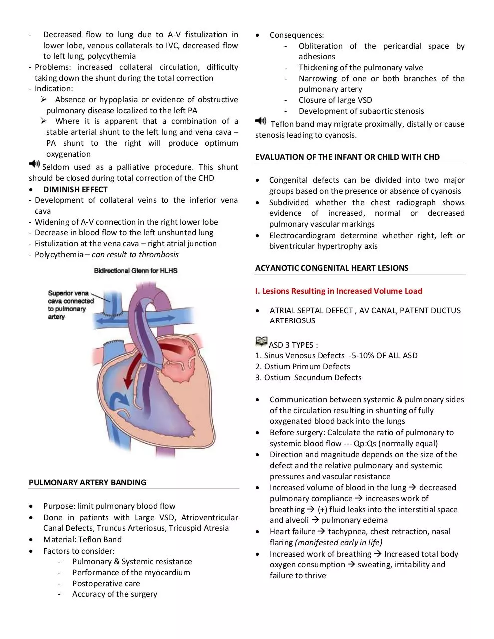
Superior Vena Cava-Right Pulmonary Artery Shunt
- first successful cavopulmonary anastomosis
Bidirectional Glenn - end-to-side RPA-to-SVC
anastomosis
Classic Glenn shunt - end-to-side right pulmonary
artery (RPA)-to-superior vena cava (SVC) anastomosis
with ligation of SVC–right atrial junction
Synthetic Interposition Grafts
- “MODIFIED BLALOCK-TAUSSIG”
- Use polytetrafluoroethylene (PTFE) graft from the
subclavian artery to the pulmonary artery
- Problems: Congestive heart failure early, shunt later
inadequate due to size restriction, kinking,
thrombosis, growth of child
Advantage: if taken down for total correction, it will
be easier to identify because it is a graft compared to
classic type
- Disadvantage: Goretex (PTFE) does not grow, as the
child grows, the graft does not grow. In later years,
there will still be decreased pulmonary blood flow.
DEFECTS REQUIRING PALLIATION (FIRST STEP BEFORE
TOTAL CORRECTION)
Tricuspid Atresia
Hypoplastic left-heart syndrome
DEFECTS THAT MAY BE PALLIATED OR REPAIRED
Ebstein’s Anomaly
Transposition of the Great Arteries
Double-Outlet Right Ventricle
Taussig-Bing Anomaly with /without pulmonary
stenosis
Tetralogy of Fallot
Ventricular Septal Defect
Atrioventricular Canal defects
Interrupted Aortic Arch
SHUNTING PROCEDURES
Cyanosis – manifestation of decreased pulmonary
blood flow
Palliation - To increase or decrease pulmonary blood
flow by creating a shunt.
Increased pulmonary blood flow – use pulmonary
artery banding and diuretics
Decreased pulmonary blood flow - create shunts
I. SYSTEMIC ARTERY – PULMONARY ARTERY SHUNTS
Subclavian Artery-Left Pulmonary Artery Shunt
- Aka “BLALOCK-TAUSSIG”
- Done in patients with decreased pulmonary blood
flow, TOF, TGA in the absence of PDA
Results to an increased pulmonary blood flow
Problems: Stenosis or thrombosis of shunt,
pulmonary hypertension (uncommon)
II. CENTRAL SHUNT
Ascending Aorta-Right Pulmonary Artery Shunt
- Aka “WATERSON”
- Problems: Kinking of pulmonary artery with
obstruction of flow to lungs, enlargement of
anastomosis, pulmonary hypertension in perfused
lung and decreased flow to contralateral lung.
Descending Aorta-Left Pulmonary Artery Shunt
- Aka “POTTS”
- Feasible only if the aorta descends on the left side
- Associated with premature closure of shunt,
enlargement of anastomosis, pulmonary
hypertension earlier
III. SYSTEMIC VEIN – PULMONARY ARTERY SHUNT
Superior Vena Cava-Right Pulmonary Artery Shunt
- Aka “GLENN SHUNT”
-
Decreased flow to lung due to A-V fistulization in
lower lobe, venous collaterals to IVC, decreased flow
to left lung, polycythemia
- Problems: increased collateral circulation, difficulty
taking down the shunt during the total correction
- Indication:
Absence or hypoplasia or evidence of obstructive
pulmonary disease localized to the left PA
Where it is apparent that a combination of a
stable arterial shunt to the left lung and vena cava –
PA shunt to the right will produce optimum
oxygenation
Seldom used as a palliative procedure. This shunt
should be closed during total correction of the CHD
DIMINISH EFFECT
- Development of collateral veins to the inferior vena
cava
- Widening of A-V connection in the right lower lobe
- Decrease in blood flow to the left unshunted lung
- Fistulization at the vena cava – right atrial junction
- Polycythemia – can result to thrombosis
Consequences:
- Obliteration of the pericardial space by
adhesions
- Thickening of the pulmonary valve
- Narrowing of one or both branches of the
pulmonary artery
- Closure of large VSD
- Development of subaortic stenosis
Teflon band may migrate proximally, distally or cause
stenosis leading to cyanosis.
EVALUATION OF THE INFANT OR CHILD WITH CHD
Congenital defects can be divided into two major
groups based on the presence or absence of cyanosis
Subdivided whether the chest radiograph shows
evidence of increased, normal or decreased
pulmonary vascular markings
Electrocardiogram determine whether right, left or
biventricular hypertrophy axis
ACYANOTIC CONGENITAL HEART LESIONS
I. Lesions Resulting in Increased Volume Load
ATRIAL SEPTAL DEFECT , AV CANAL, PATENT DUCTUS
ARTERIOSUS
ASD 3 TYPES :
1. Sinus Venosus Defects -5-10% OF ALL ASD
2. Ostium Primum Defects
3. Ostium Secundum Defects
PULMONARY ARTERY BANDING
Purpose: limit pulmonary blood flow
Done in patients with Large VSD, Atrioventricular
Canal Defects, Truncus Arteriosus, Tricuspid Atresia
Material: Teflon Band
Factors to consider:
- Pulmonary & Systemic resistance
- Performance of the myocardium
- Postoperative care
- Accuracy of the surgery
Communication between systemic & pulmonary sides
of the circulation resulting in shunting of fully
oxygenated blood back into the lungs
Before surgery: Calculate the ratio of pulmonary to
systemic blood flow --- Qp:Qs (normally equal)
Direction and magnitude depends on the size of the
defect and the relative pulmonary and systemic
pressures and vascular resistance
Increased volume of blood in the lung decreased
pulmonary compliance increases work of
breathing (+) fluid leaks into the interstitial space
and alveoli pulmonary edema
Heart failure tachypnea, chest retraction, nasal
flaring (manifested early in life)
Increased work of breathing Increased total body
oxygen consumption sweating, irritability and
failure to thrive
Remember that severe pulmonary hypertension is a
contraindication to definitive surgery in patients with
CHD
-
II. Lesions Resulting in Increased Pressure Load –
obstruction to normal blood flow
Obstruction to ventricular outflow:
PULMONARY STENOSIS
- Critical Pulmonary Stenosis (
very small
orifice) in newborn presents as right-sided heart
failure hepatomegaly, peripheral edema,
cyanosis (shunting across foramen ovale)
AORTIC STENOSIS
- Critical Aortic Stenosis(very small orifice) in
newborn presents as left-sided heart failure
pulmonary edema, poor perfusion, and rightsided heart failure
COARCTATION OF THE AORTA
Obstruction to ventricular inflow:
TRICUSPID STENOSIS
MITRAL STENOSIS
- As a congenital defect, it is seldom seen
- More often a complication of rheumatic heart
disease
CYANOTIC CONGENITAL HEART DISEASE
I. DECREASED PULMONARY BLOOD FLOW
- Obstruction to pulmonary blood flow and a pathway
by which systemic venous blood can shunt from right
to left
TRICUSPID ATRESIA
TETRALOGY OF FALLOT
II. INCREASED PULMONARY BLOOD FLOW
- Cyanosis caused by either abnormal ventriculararterial connections or by total mixing of systemic
venous and pulmonary venous blood within the heart
TOTAL ANOMALOUS PULMONARY VENOUS
RETURN (TAPVR)
TRUNCUS ARTERIOSUS
TETRALOGY OF FALLOT
VENTRICULAR SEPTAL DEFECT
Most common: 25% of CHD
Defects occur in any portion if ventricular septum; the
majority are of the membranous type
Location: Anterior to the septal leaflet of tricuspid
valve
Between supraventricularis and papillary
muscle of conus
Location: Superior to crista supraventricularis
- Just beneath the pulmonary valve and may
impinge on an aortic sinus
- Midportion or apical region of the ventricular
septum – muscular type – single or
multiple(catheterization is indicated due to
multiple VSD)
DETERMINANT OF THE SIZE OF SHUNT
Size of VSD
Level of PVR compared with SVR, O2 saturation
Small, <0.5 cm – aka restrictive
- RV pressure is normal
- No indication for immediate medical intervention
>1.0 cm – aka nonrestrictive
- RV & LV pressures are equalized
Pulmonic Vascular Resistance (PVR): Systemic Vasuclar
Resistance (SVR) = 1:1
- The shunt becomes bidirectional
- Signs of heart failure abate and patient becomes
cyanotic
Small Shunt
- Qp:Qs <1.75
- Cardiac chambers not enlarged
- Pulmonary vascular bed are normal
Large Shunt
- Qp:Qs >2:1
- Left atrial & ventricular volume overload occurs
- Enlarged main pulmonary artery, left atrium, left
ventricle
CLINICAL MANIFESTATION
Small VSD
- Asymptomatic
- Found on routine physical exam
Large VSD
- Excessive pulmonary blood flow and pulmonary
hypertension
- Dyspnea, feeding difficulties, poor growth,
profuse perspiration, recurrent pulmonary
infection, cardiac failure in early infancy
- Duskiness during infections or crying
- Prominence of left precordium and palpable
parasternal lift
- Laterally displaced apical impulse
- Increase pulmonic component of 2nd sound
o Pulmonary hypertension
- Less harsh holosystolic murmur
-
Mid-diastolic low-pitched rumble at the apex –
because increased blood flow across mitral valve
DIAGNOSIS
Small VSD
- Chest radiograph – normal
- ECG – normal
Large VSD
Chest radiograph
- Cardiomegaly
- Increased pulmonary markings
ECG
- Biventricular hypertrophy
2D Echocardiogram
- Position & size of VSD
- Estimate shunt size
- Associated lesions
- Calculate pressure gradient
Cardiac Catheterization
Important to identify multiple VSD
- Complications: premature rupture of balloon, air
embolism
PROGNOSIS
Small defect – 30-35% spontaneous closure
- Small muscular defect are more likely (80%) to
close than membranous (35%)
Large defect – less common to close
Advise surgical intervention at an early age to prevent
heart failure. If large defect, right away advise VSD
closure. If small defect, observe and monitor. Advise
not to become hypoxemic because VSD won’t close.
TREATMENT
Small VSD
- No restrictions of physical activity
- Surgery not recommended
- Protection against infective endocarditis
Patient should have antibiotics before any
interventionand dental clearance.
Indications for surgery:
- Large defects with clinical symptoms and failure
to thrive which cannot be controlled medically
- Infants between 6-12 months with large defects,
with pulmonary hypertension
- Patients older than 24 months with Qp:Qs > 2:1
- Supracritical VSD – high risk of Aortic
Insufficiency because of proximity.
Sources: slides, Schwartz, audio
By M. Prado, R. Gabor, K. Carvajal, N. Sameon
Download #1 Surgical Management of Congenital Heart Disease
#1 Surgical Management of Congenital Heart Disease.pdf (PDF, 568.93 KB)
Download PDF
Share this file on social networks
Link to this page
Permanent link
Use the permanent link to the download page to share your document on Facebook, Twitter, LinkedIn, or directly with a contact by e-Mail, Messenger, Whatsapp, Line..
Short link
Use the short link to share your document on Twitter or by text message (SMS)
HTML Code
Copy the following HTML code to share your document on a Website or Blog
QR Code to this page

This file has been shared publicly by a user of PDF Archive.
Document ID: 0000134943.