PBFD (PDF)
File information
Author: s.yamashkina
This PDF 1.5 document has been generated by Microsoft® Word 2010, and has been sent on pdf-archive.com on 22/07/2014 at 17:20, from IP address 220.142.x.x.
The current document download page has been viewed 7991 times.
File size: 612.85 KB (42 pages).
Privacy: public file


File preview
1
http://www.avianbiotech.com
Psittacine Beak and Feather
Disease (PBFD)
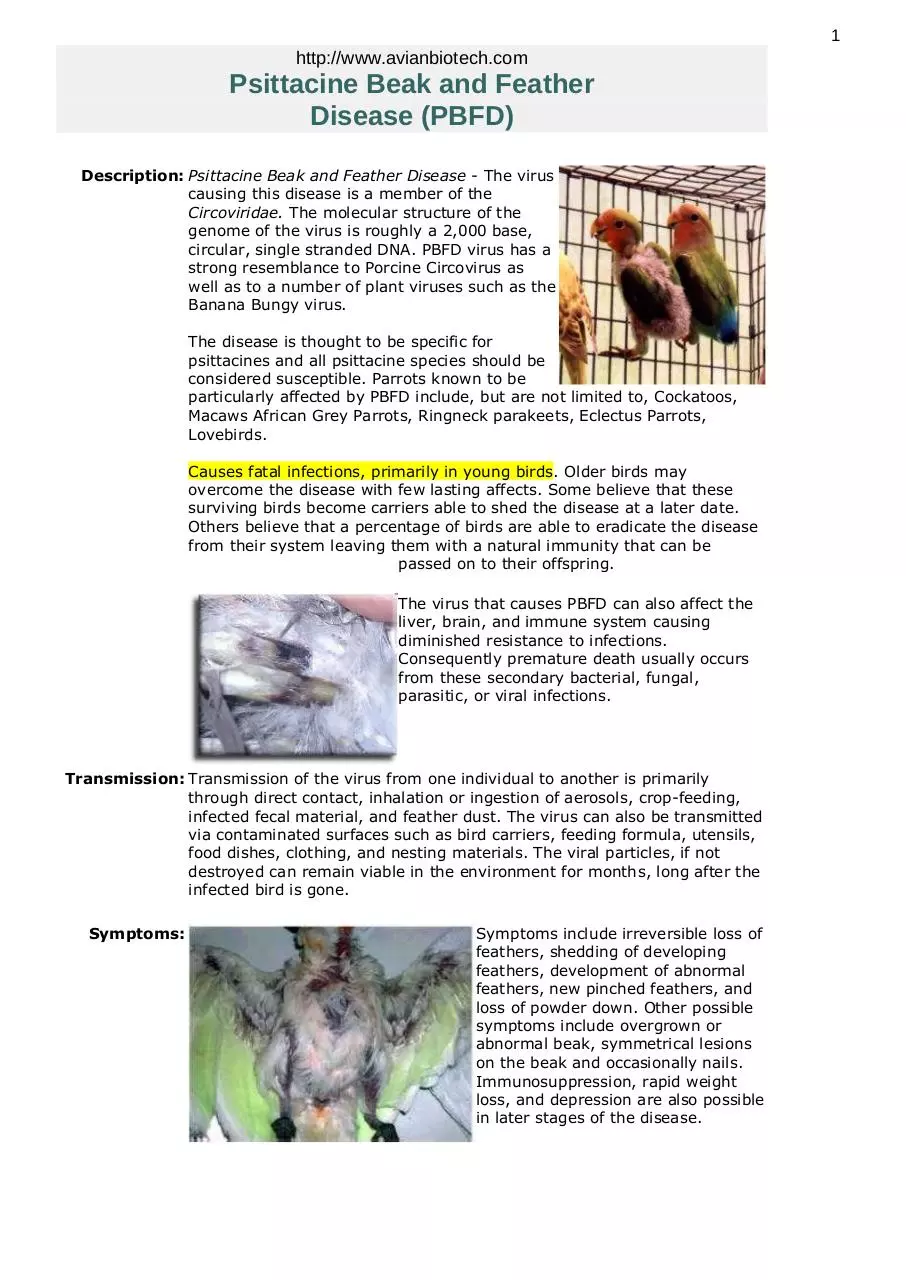
Description: Psittacine Beak and Feather Disease - The virus
causing this disease is a member of the
Circoviridae. The molecular structure of the
genome of the virus is roughly a 2,000 base,
circular, single stranded DNA. PBFD virus has a
strong resemblance to Porcine Circovirus as
well as to a number of plant viruses such as the
Banana Bungy virus.
The disease is thought to be specific for
psittacines and all psittacine species should be
considered susceptible. Parrots known to be
particularly affected by PBFD include, but are not limited to, Cockatoos,
Macaws African Grey Parrots, Ringneck parakeets, Eclectus Parrots,
Lovebirds.
Causes fatal infections, primarily in young birds. Older birds may
overcome the disease with few lasting affects. Some believe that these
surviving birds become carriers able to shed the disease at a later date.
Others believe that a percentage of birds are able to eradicate the disease
from their system leaving them with a natural immunity that can be
passed on to their offspring.
The virus that causes PBFD can also affect the
liver, brain, and immune system causing
diminished resistance to infections.
Consequently premature death usually occurs
from these secondary bacterial, fungal,
parasitic, or viral infections.
Transmission: Transmission of the virus from one individual to another is primarily
through direct contact, inhalation or ingestion of aerosols, crop-feeding,
infected fecal material, and feather dust. The virus can also be transmitted
via contaminated surfaces such as bird carriers, feeding formula, utensils,
food dishes, clothing, and nesting materials. The viral particles, if not
destroyed can remain viable in the environment for months, long after the
infected bird is gone.
Symptoms:
Symptoms include irreversible loss of
feathers, shedding of developing
feathers, development of abnormal
feathers, new pinched feathers, and
loss of powder down. Other possible
symptoms include overgrown or
abnormal beak, symmetrical lesions
on the beak and occasionally nails.
Immunosuppression, rapid weight
loss, and depression are also possible
in later stages of the disease.
2
Secondary viral, fungal, bacterial or parasitic
infections often occurs as a result of
diminished immunity caused by a PBFD viral
infection. Additional symptoms not
mentioned above including elevated white
cell counts are generally due to secondary
infections and may not be directly related to
PBFD virus infections.
Prevention: Strict isolation of all diseased birds to halt the spread of the disease. DNA
testing of all birds of susceptible species to rule out latent infection. DNA
testing of aviary equipment and environment to test for possible
contamination.
Treatment: No known treatment. Experimental vaccines are being developed.
Diagnosis: Skin biopsy, surgical biopsy of feather and shaft, or PCR testing of blood,
swab, and feather samples.
PBFD should be considered in any bird suffering from abnormal feather
loss or development. A biopsy of the abnormal feathers including the
calimus (shaft) of the feather can be examined for signs of virus. However,
since the PBFD virus does not affect all feathers simultaneously this
method of evaluating a sample may have a high degree of error.
Additionally, birds with PBFD can have normal feathers and the PCR test is
the most effective method available for detecting the virus in birds before
feather lesions develop.
Some birds infected with the virus, test positive, but never show clinical
signs. Other birds which test positive may develop an immune response
sufficient enough to fight off the infection and test negative after 30-90
days. Therefore, it is recommended to re-test all PBFD positive birds 60-90
days after the initial testing was completed. If the second sample remains
positive, the bird should be considered permanently infected and can be
expected to show clinical symptoms of the disease.
Sample: To test an individual bird a whole blood sample is recommended in
conjunction with a cloacal swab or feathers (especially abnormal or
suspicious-looking feathers) when possible. If
the sample tests
positive the bird should be placed in
quarantine and re-tested after 4-6 weeks. If
the bird tests negative the second time a third
test after 4-6 weeks is recommended.
Post-mortem samples include liver, spleen,
kidney, feather samples in a sterile container;
postmortem swabs may also be submitted.
Environmental testing using swabs of aviaries, countertops, fans, airfilters, nest-boxes, etc. is extremely effective in determining the presence
of PBFD DNA in the environment.
*It is recommenced to submit both a whole blood and cloacal swab sample
for analysis when possible.
Handling: Prior to shipping samples should be stored at 4 C. (refrigerator). Samples
must be shipped in a padded envelope or box. Samples may be sent by
regular mail, but overnight is recommended.
3
Avian Biotech International
1336 Timberlane Road · Tallahassee, FL 32312-1766
850-386-1145 or 800-514-9672 (Office) 850-386-1146 (Fax)
Copyright © 1995-2005 Animal Genetics, Inc. All rights reserved.
Avian Biotech and Avian Connection are ™ of Animal Genetics, Inc.
4
5
http://www.peteducation.com/article.cfm?cls=15&cat=1829&articleid=2592
Psittacine Beak and Feather Disease (PBFD)
Holly Nash, DVM, MS
Veterinary Services Department, Drs. Foster & Smith, Inc.
Psittacine Beak and Feather Disease (PBFD) is a contagious, fatal viral disease that affects the beak,
feathers, and immune system of birds belonging to the Psittacidae family. It was first recognized in 1975
by veterinarians in Australia, where the disease affects wild birds. Although birds showing signs of disease
usually die, it is common for birds to be exposed to the virus, develop a mild infection, and recover.
What birds are at risk for PBFD?
PBFD has been diagnosed in over 40 species of psittacines, including South American parrots. Although all
members of this family appear to be susceptible, PBFD is seen more often in cockatoos. Eclectus parrots,
lovebirds, budgies, and African grey parrots are also affected. Younger birds are more commonly affected,
especially with the acute form of the disease. Most birds affected are under 2 years of age.
What causes PBFD?
PBFD is caused by a DNA virus that affects the cells of the immune system and those that produce the beak
and feathers. The virus is a circovirus, which is one of the smallest viruses known to cause disease. A
similar virus affects doves and other birds.
How is the virus that causes PBFD transmitted?
PBFD is extremely contagious. Large amounts of the virus, which can become airborne, are found in the
droppings, contents of the crop, and the feather dust of infected birds. The feather dust is easily dispersed
and can contaminate food, water, cages, clothing, and other areas of the environment. The virus is thought
to be transmitted by inhalation or ingestion of the virus. It has been suggested that the virus may be
transmitted in utero from the female bird to the egg.
The incubation period (time between exposure to the virus and the development of signs) can be as short
as 3-4 weeks, or up to several years, depending upon the amount of virus transmitted, the age of the bird,
the stage of feather development, and the health of the bird's immune system.
What are the signs of PBFD?
There are both acute and chronic forms of the disease.
Peracute/Acute Form: The peracute and acute forms most commonly occur in very young birds, and may begin
with signs unrelated to the beak or feathers. Affected birds are often depressed and regurgitate due to crop
stasis. They may develop a diarrhea-causing enteritis or pneumonia, and die without displaying any lesions of
the feathers or beak. This is often called the peracute form of the disease. In the acute form, juveniles
losing their down and developing feathers may have lesions on the feathers, including circular bands
around the feathers which constrict its base. These feathers are often loose, break easily, may bleed, and
are very painful.
Common Signs of Psittacine Beak & Feather Disease
Acute Form
Depression
Regurgitation and diarrhea
Loss of appetite and weight
Abnormal feather development
Death
Chronic Form
Loss of feather dust and powder
Abnormal feather development
Abnormal growth and deformities of the beak
Necrotic beak and oral lesions
Secondary infections
Death in months to years
Chronic Form: In the chronic form of PBFD, which is more common in older birds, the powder-down feathers
are often the first feathers affected. The feathers are fragile and fracture easily, have constricting bands,
may hemorrhage, and may be discolored, deformed, or curled. As the feather follicles are damaged, the bird
will soon be unable to replace feathers, and the primary, secondary, tail, and crest feathers are lost. Bare
skin is exposed, and the normal feather dust is not found on the body or the beak, where it normally
accumulates due to preening. Feather abnormalities, often termed "dystrophic feathers," may not appear until
the first molt after infection, which could be a period up to 6 months.
6
The beak may develop irregular sunken areas. Brown necrotic areas may be found inside the upper beak,
and the beak may elongate, become deformed, and fracture. Secondary beak and oral infections often
occur. In some birds, the nails can also be deformed or slough.
Mucus in the droppings, or a green tint to the droppings may occur. In some birds, the
liver will be affected, and liver failure may be the cause of death.
Birds with the chronic form of the disease may live for months to years before dying of a
secondary infection. This long period of illness in which the bird may be featherless, and
gradually weakens can be very emotionally difficult for owners.
How is PBFD diagnosed?
The review of the medical history, presence of clinical signs, and observations during the physical exam
support the diagnosis of PBFD. Other conditions such as nutritional deficiencies, infection with
polyomavirus (causes budgerigar fledgling disease and other diseases of psittacines), hormonal
abnormalities, and drug reactions can cause lesions on the feathers similar to PBFD. Histopathology
(microscopic examinations of biopsies) can confirm the diagnosis. Affected cells will have abnormalities in
their nuclei, called "basophilic intranuclear inclusion bodies." The diagnosis may also be confirmed by a PCR
(polymerase chain reaction) test on whole blood or biopsy samples from the affected bird. The test detects
the presence of the virus. This test may also be used on swabs of surfaces in the environment to detect
contamination.
False positive and false negative test results can occur. For example, infected airborne cells could
contaminate a sample and cause a false positive result. Healthy birds with a positive test result should be
retested after 90 days. If they still have positive test results, they should be considered carriers of the
virus. If the retest is negative, the bird may have eliminated the virus, and become immune.
False negative results may occur if too much anticoagulant is present in the sample, an extremely high
number of viral particles are present and interfere with the test, or there are an insufficient number of
infected white blood cells in the sample.
How is PBFD treated?
There is no specific treatment for PBFD. Supportive care including good nutrition, supplementary heat
(incubator), beak trimming, and treatment of secondary infections can be offered. The disease, however, is
progressive, and very few birds recover. Euthanasia may need to be considered for birds with severe
and/or painful signs. Birds who die a natural death usually succumb to a secondary bacterial, fungal, or
viral infection despite treatment, since their immune systems have been critically suppressed. Most birds
die within 6 months to 2 years of developing the disease.
How is PBFD prevented and controlled?
Birds should be purchased from suppliers with disease-free birds. New birds coming into facilities should be
quarantined and tested. Repeat testing in 3-4 weeks to allow for the incubation period is recommended.
Infected birds should be isolated and removed from breeding programs. Juvenile birds should be housed
separately from adults. Bird owners need to understand that if they handle other peoples' birds, it may be
possible for them to bring the virus into their home and infect their birds. Good hygiene and sanitation
should be used. The susceptibility of the virus to disinfectants is unknown. Disinfectants which are known
to be effective against parvoviruses are probably the best choice.
In Australia, a killed vaccine has been developed which can protect unexposed birds; it can cause more severe
disease in birds already showing signs of PBFD. Birds should be vaccinated as young as possible, as soon
as 14 days of age. The vaccine should be boostered after one month, and breeding birds should be
vaccinated one month prior to breeding.
References and Further Reading
Altman, RB; Clubb, SL; Dorrestein, GM; Quesenberry, K. Avian Medicine and Surgery. W.B. Saunders Co. Philadelphia, PA; 1997.
Raidal, SR. http://wwwvet.murdoch.edu.au/caf/pbfd.htm. Murdoch University. Perth, Western Australia.
Rupley, AE. Manual of Avian Practice. W.B. Saunders Co. Philadelphia, PA; 1997.
7
8
http://www.vetark.co.uk/pbfd.html
This dreadful disease is caused by a circovirus. It has a wide species range although it appears to be a
natural virus infection of cockatoos in Australasia where it occurs in wild flocks. It has been known in wild
cockatoos in Australia for many years and recently Ducorps cockatoos from the Solomon Islands have
been round to be infected. Old world parrots show the infection most commonly. In the US eclectus and
cockatoos led African greys. New World parrots such as Amazons and macaws showed less of the
disease. Smaller species such as lovebirds, cockatiels and parakeets also showed the infection very
commonly.African greys as an unnatural host seem particularly acutely affected, young birds may simply die or
develop feather loss first, others may develop red feathering (seen in wild birds also unrelated to PBFD).
Feather colour changes are also reported in Vasa parrots. It causes typical French Moult signs in
budgies, and similar signs in lovebirds and ringnecks. A few species have been reported to eliminate
infection and recover, this seems commoner in lovebirds than any other species.Variable levels of feather loss are seen, in some birds it may develop slowly with only a few abnormal
feathers each moult. Rapidly growing feathers are affected first, eg. powder downs in african greys and
cockatoos, losing their natural dust they often develop an untidy greasy plumage and shiny beaks. It may
be seen at the first formation of feathers to replace down. Sudden loss or deformities of feathers, often
blood in the sheath. It may also develop in adult birds at subsequent moults. Perhaps a few affected
feathers each time.The virus is found in feather dust, faeces and in crop fluids. It is believed to spread through the egg.
It has been reported that birds may simply die of either disease without showing signs. Liver or kidney
swabs clipped off and dropped into carrier medium may be sent from probing if these diseases are
suspected. PBFD tests PCR testing is very sensitive. Contamination by virus from other positive birds,
whether dead or alive will make a sample positive. The test finds virus if it is there - from any source.
In just the same way that your sample may be contaminated from via environment (from an unsuspected
carrier) it is not uncommon for chicks to show severe problems yet parents are negative. These chicks
are often being infected from a contaminated environment.
We cannot control potential contamination at the time of collection so ensuring the sample is not
contaminated is your responsibility. To avoid contamination of the sample with PBFD virus from the
environment (originating from other birds) it is essential to thoroughly clean the birds nail area. We
recommend Ark-Klens (from VETARK) for this. RELYING ON DISINFECTION IS NOT SUFFICIENT.
CLEANSING IS VITAL. Dead virus will give as strong a positive reaction as live virus. Alternatively have
your veterinary surgeon collect the sample direct from a vein by venipuncture. Birds become immunosuppressed and may die from other diseases.
You can now use the standard DNA collection kits to take blood from parrots for PBFD testing.
Alternatively a veterinary surgeon will collect the sample directly from a vein using a syringe and needle
(venipuncture). Feather pulp from abnormal feathers is also a potential source of virus. This is squeezed
straight into our collection tubes. Because birds with a serious degree of feather lesions may be
immunosuppressed, and because this can hide the virus we recommend that such birds be sampled by
collecting blood and feather pulp samples in the same tube. Do not simply send us dried feathers - they
are a poor sample for reliable testing - so as a policy we don't test them.
Please use or at the very least liaise with your avian vet. Interpreting what the test means to you and
your birds and what if any action is required in your situation needs veterinary input. We can assist your
vet but we cannot get involved with cases directly.
What does a positive test result mean ?
9
A positive PBFD test result means that the the PCR test detected PBFD DNA in the sample. A positive
result from a bird with feather abnormalities suggests strongly that the bird has an active infection.
A positive result from a bird with no feather problems may mean either that the bird is a carrier or that it
has been recently exposed to the virus. In these cases we recommend re-testing in 90 days. We also
recommend that the second sample is collected by venipuncture to ensure that contamination does not
occur. The majority of birds which are merely exposed will mount an immune response and eliminate the
infection.
Those still positive at the 90 day test should be considered carriers. One day they are likely to show the
disease, and be potentially infectious.
Various uses of the disease tests
to test clinically suspect birds
to examine material from post mortem examinations of dead birds
to check collections for carriers and to look for in-contacts
to test 'new birds' at pre- or post-purchase veterinary health checks eg. as pets or before entering
breeding collections
to test birds in the pet shop
Download PBFD
PBFD.pdf (PDF, 612.85 KB)
Download PDF
Share this file on social networks
Link to this page
Permanent link
Use the permanent link to the download page to share your document on Facebook, Twitter, LinkedIn, or directly with a contact by e-Mail, Messenger, Whatsapp, Line..
Short link
Use the short link to share your document on Twitter or by text message (SMS)
HTML Code
Copy the following HTML code to share your document on a Website or Blog
QR Code to this page

This file has been shared publicly by a user of PDF Archive.
Document ID: 0000175577.