JDIT 2014 1125 010 (PDF)
File information
Title:
Author: Sean
This PDF 1.5 document has been generated by Microsoft® Word 2010, and has been sent on pdf-archive.com on 30/05/2017 at 00:32, from IP address 90.218.x.x.
The current document download page has been viewed 442 times.
File size: 522.87 KB (20 pages).
Privacy: public file


File preview
Journal of Diagnostic Imaging in Therapy. 2014; 1(1):137-156
Mansi et al.
Open Medscience
Peer-Reviewed Open Access
JOURNAL OF DIAGNOSTIC IMAGING IN THERAPY
Journal homepage: www.openmedscience.com
Review Article
Basic Premises to Molecular Imaging and Radionuclide
Therapy – Part 1
Luigi Mansi1,*, Sean L Kitson2, Vincenzo Cuccurullo1, Andrea Ciarmiello3
1
Nuclear Medicine Unit Department of Clinical and Experimental Internistic ‘F. Magrassi, A.
Lanzara’- Seconda Università di Napoli Naples, Italy
2
Department of Biocatalysis and Isotope Chemistry, Almac, 20 Seagoe Industrial Estate, Craigavon,
BT63 5QD, United Kingdom
3
Nuclear Medicine Department, S. Andrea Hospital, La Spezia, Italy
*Author to whom correspondence should be addressed:
Luigi Mansi, M.D.
luigi.mansi@unina2.it
Abstract
This manuscript is a complementary article to an accompanying paper, published in a forthcoming
issue, which will give an overview on the central role of chelation in labelling radiocompounds; either
for imaging and/or for radionuclide therapy. In order to facilitate a better understanding of the
importance of Chelator-Based Imaging & Therapy, we will briefly discuss in this publication - which
is partially intended as an introduction to the second paper - which contains the major basic principles
of molecular imaging and radionuclide therapy. Although these issues are of interest in the general
field of Nuclear Medicine; since the chelation process involves the labelling with radiometals; we aim
to highlight examples of this category in this paper which concern this class of nuclides in this
particular issue.
ISSN: 2057-3782 (Online)
http://dx.doi.org/10.17229/jdit.2014-1125-010
137
Journal of Diagnostic Imaging in Therapy. 2014; 1(1):137-156
Mansi et al.
Keywords: molecular imaging; radionuclide therapy; radiotracers; chelation; radiometals; radiohalogens; SPECT imaging; PET imaging
1. Introduction
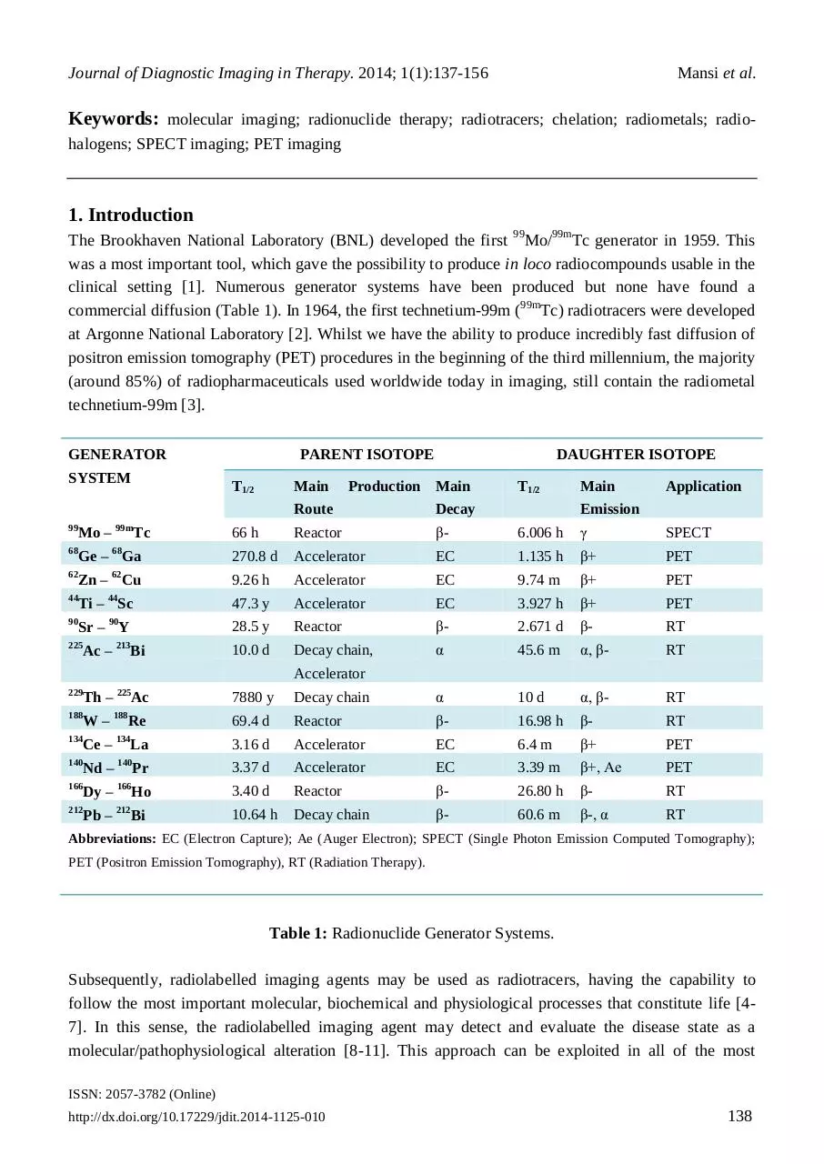
The Brookhaven National Laboratory (BNL) developed the first 99Mo/99mTc generator in 1959. This
was a most important tool, which gave the possibility to produce in loco radiocompounds usable in the
clinical setting [1]. Numerous generator systems have been produced but none have found a
commercial diffusion (Table 1). In 1964, the first technetium-99m (99mTc) radiotracers were developed
at Argonne National Laboratory [2]. Whilst we have the ability to produce incredibly fast diffusion of
positron emission tomography (PET) procedures in the beginning of the third millennium, the majority
(around 85%) of radiopharmaceuticals used worldwide today in imaging, still contain the radiometal
technetium-99m [3].
GENERATOR
SYSTEM
PARENT ISOTOPE
T1/2
Main
DAUGHTER ISOTOPE
Production Main
T1/2
Main
Application
Route
Decay
66 h
Reactor
β-
6.006 h
γ
SPECT
Ge – Ga
270.8 d
Accelerator
EC
1.135 h
β+
PET
Zn – Cu
9.26 h
Accelerator
EC
9.74 m
β+
PET
Ti – Sc
47.3 y
Accelerator
EC
3.927 h
β+
PET
Sr – Y
28.5 y
Reactor
β-
2.671 d
β-
RT
10.0 d
Decay chain,
α
45.6 m
α, β-
RT
99
Mo –
68
62
44
90
99m
Tc
68
62
44
90
225
Ac –
213
229
Th –
225
188
W–
188
134
Ce –
134
140
Nd –
140
166
Dy –
166
212
Pb –
212
Bi
Emission
Accelerator
Ac
7880 y
Decay chain
α
10 d
α, β-
RT
Re
69.4 d
Reactor
β-
16.98 h
β-
RT
3.16 d
Accelerator
EC
6.4 m
β+
PET
Pr
3.37 d
Accelerator
EC
3.39 m
β+, Ae
PET
Ho
3.40 d
Reactor
β-
26.80 h
β-
RT
Bi
10.64 h
Decay chain
β-
60.6 m
β-, α
RT
La
Abbreviations: EC (Electron Capture); Ae (Auger Electron); SPECT (Single Photon Emission Computed Tomography);
PET (Positron Emission Tomography), RT (Radiation Therapy).
Table 1: Radionuclide Generator Systems.
Subsequently, radiolabelled imaging agents may be used as radiotracers, having the capability to
follow the most important molecular, biochemical and physiological processes that constitute life [47]. In this sense, the radiolabelled imaging agent may detect and evaluate the disease state as a
molecular/pathophysiological alteration [8-11]. This approach can be exploited in all of the most
ISSN: 2057-3782 (Online)
http://dx.doi.org/10.17229/jdit.2014-1125-010
138
Journal of Diagnostic Imaging in Therapy. 2014; 1(1):137-156
Mansi et al.
significant pathological fields with the potential to detect and/or biologically characterize issues such
as organ function, tumour malignancy, blood flow alterations, metabolic processes, in vivo receptor
distribution and so forth [12-15].
In addition, as well as being used as a diagnostic tool, radiopharmaceuticals also find applications in
therapy - notably in the treatment of cancer. However, the clinical application in benign diseases are
widely diffuse [16,17]. The Imaging Periodic Table (Table 2) gives an overview of the types of
radiation emitted from the various radionuclides.
H
Li
Alpha emitters α; Beta (electron) emitters β-;
Positron emitters β+; Gamma emitters γ;
Auger electrons Ae
Be
Na
Mg
K
Ca
Sc
Ti
V
Cr
Mn Fe
γ
βRb
Sr
Y
Ba
La
Co
Ni
β+ β+
Zr
Nb
Mo Tc
β- β+
Cs
Hf
Ta
W
Ra
Ac
α
α
Ce
Pr
Nd
Ru
Rh
Pd
γ
γ
β-
γ
β-
β- γ
β-
Re
Os
Ir
Pt
Pa
U
Pm Sm Eu
Np
Pu
Ag
Au
Gd
Tb
Dy
Al
Si
Cf
N
O
Ga
Ge
P
F
Ne
β+
S
Cl
Ar
Se
Br
Kr
βZn
ββ+
γ
Cd
In
As
β-
Sn
Sb
Ae
Te
γ
Hg TI
Ho
Er
Es
I
Xe
γ
βAe
Pb
Bi
Po
β- α
Ae
β- βAm Cm Bk
C
β+ β+
Ae
γ
βTh
Cu
B
ββ+
γ
γ
βFr
He
The Imaging Periodic Table
Tm Yb
At
Rn
α
Lu
β- βFm Md No
Lr
Table 2: The Imaging Periodic Table.
When a radiopharmaceutical is introduced into the body, it localises in particular tissue(s) on the basis
of a distribution determined by its action as a radiotracer of a biological process. The extent to which
this happens is dependent either on the radiocompound and/or on pathophysiological characteristics of
the tissue(s) [18]. A specific radiopharmaceutical may be concentrated at the site of a lesion such as a
neoplasm; an infective process and/or of a particularly normal tissue. These processes follow
molecular events generally typical for uptake within a tissue(s), although rarely pathognomonic [19].
ISSN: 2057-3782 (Online)
http://dx.doi.org/10.17229/jdit.2014-1125-010
139
Journal of Diagnostic Imaging in Therapy. 2014; 1(1):137-156
Mansi et al.
The patient can then be scanned in order to image the radioactivity: this generates a real-time imagine
of what is happening in the body [20]. A significant advantage of this method over other imaging
techniques is that the ponderal amount of radioactive compounds is very small - often nanomolar
quantities or less [21,22]. This is a major advantage for use in molecular imaging with respect to the
two main systems currently in use such as computed tomography (CT) and magnetic resonance
imaging (MRI), when performed with contrast media.
In fact, gadolinium-containing agents or iodinated contrast media are used to enhance and/or modify
the image. These imaging agent are administered in millimolar quantities and for these reasons they
cannot be used to image many of the most important molecular processes as neurotransmission,
metabolism etc. Subsequently, these processes can only be studied using tracers in the order of
micro/nanomoles.
Conversely, the possibility of molecular imaging being in direct competition with nuclear medicine by
using optical imaging (OI) has the capability to trace all of the most crucial biological events.
Nevertheless, being optical imaging based, using light as the source of the image means that it can not
be accurately used in vivo in humans to analyse deep organs. This is because light photons are not able
to pass through the tissue matter [23].
2. Pathophysiological Premises to Nuclear Medicine
Nuclear Medicine is the field in diagnostic imaging where the value of the image is less dependent on
the scanner. The radiotracer is used in the clinical context for radionuclide studies. In order to
understand this particular concept, a good precedent can derive from the study used for detecting
Meckel’s diverticulum which is the most frequent cause of bleeding in paediatrics. Nevertheless, at
this time the most reliable diagnostic procedure is the planar scintigraphy used with 99mTcpertechnetate. The major advantage of planar scintigraphy is its commercial cost effectiveness per
patient over the remaining procedures which include PET, SPECT and other radiological techniques
such as ultrasound (US), CT and MRI [24].
Although the spatial resolution of a standard gamma camera is typically more than 1 cm, the
scintigraphy is able to detect the area of gastric ectopic mucosa at level of the bowel. Also, it may
establish the pathophysiological cause of the bleeding. In fact, the radiotracer used for example 99mTcpertechnetate, is biologically analogous of iodine. The imaging agent 99mTc-pertechnetate concentrates
in thyroid, salivary gland, choroid plexus and gastric mucosa. This behaviour leads to a diagnosis
based on a radiotracer’s concentration at the level of the ectopic gastric tissue. This allows diagnosis
which is not normally feasible by morphostructural techniques not with standing their higher spatial
resolution.
This means that in presence of an increased uptake of the radiotracer which would have a favourable
lesion/background concentration rate, the information is substantially independent of the spatial
resolution from the diagnostic instrument.
ISSN: 2057-3782 (Online)
http://dx.doi.org/10.17229/jdit.2014-1125-010
140
Journal of Diagnostic Imaging in Therapy. 2014; 1(1):137-156
Mansi et al.
This peculiarity is associated with the so called functional studies, for example in nuclear medicine
functional techniques such as Rx, CT, MRI and US, with or without contrast media; in which the
image is dependent on a pathophysiological premise, and is not only expression by differences in
density as in morphostructural studies.
In other words, these functional studies represent the ‘living function’: normal (physiology) or altered
(pathophysiology). Conversely, morphostructural techniques are used to evaluate ‘anatomy’ and
‘pathological data’, i.e. static information having substantially the same content in the living body
and/or in a non-living subject.
Furthermore, these functional alterations precede the establishment of a pathological change, as in the
non-living subject. Therefore, it is possible to acquire functional procedures with an earlier diagnosis,
being achievable original information being more strictly connected with prognosis and therapy [25].
3. Gamma Emitters and SPECT Imaging
There is a broad spectrum of radionuclides and between them a large number of radiometals are
currently being used for medical imaging and therapy. They have different mechanisms of radioactive
decay, making them suitable for particular applications [26]. The most commonly used diagnostic tool
involves radiometals which emit gamma rays. The gamma rays are detected using a gamma camera
[27]. Typically, this system is based on a detector capturing the rays with a sodium iodide crystal; light
photons obtained by ‘scintillation’ are then transformed into electric signals, amplified and elaborated
in order to create an image.
Figure 1: A schematic diagram of SPECT Imaging.
ISSN: 2057-3782 (Online)
http://dx.doi.org/10.17229/jdit.2014-1125-010
141
Journal of Diagnostic Imaging in Therapy. 2014; 1(1):137-156
Mansi et al.
A similar process is maintained when using more modern detecting systems, as those using solid
detectors [28]. Historically created as analogical machines; at the present all gamma cameras are fully
digitalized. The final image may be either reconstructed in a single plane, to obtain the so called static
or sequential ‘scintigraphy’, or three-dimensionally, using a single photon emission computed
tomography (SPECT) being able to produce a tomoscintigraphy with gamma emitters, i.e. a SPECT
image (Figure 1).
A further significant improvement has been achieved with the diffusion of the so called hybrid
systems; constructed in the same gantry with two (or exceptionally three) different imaging tools.
Typically, a nuclear medicine machine [29] such as (SPECT, PET) combined with a CT scanner [30].
More recently, PET/MRI hybrid systems have been commercialized [31,32], although still in the
experimental preclinical field SPECT/MRI machines are still being commercially developed [33].
The major advantage of hybrid machines is the possibility of integrating the functional image obtained
by radionuclide procedures with morphostructural data acquired by CT (or MRI). In this instance a
significant improvement in diagnostic accuracy is reached; together with further and/or better
capabilities, as those connected with a utilization of the hybrid system as a guide for a biopsy or in the
better biological definition of a target for radiotherapy. It has to be pointed out that at present all the
CT scanners included in a hybrid machine are diagnostic multi-slice CT (MSCT) [34].
Scintigraphy and SPECT are the final result of a signal starting from radionuclides that emit gamma
photons of a given energy [35]. In Table 3 are represented the actually more frequently used gamma
emitters.
It is noted that, between them, the most diffuse is certainly 99mTc, which utilized in more than 95% of
the examinations performed with a gamma camera. The use of 67Ga, as citrate [36], is nearly
disappeared, having being completely overwhelmed by the clinic explosion of PET with 18F-fluorodeoxyglucose (18FDG).
The diagnostic use of iodine-131, which emits beta radiation [37] is used in radionuclide therapy and is
almost only limited to the whole body evaluation performed in the follow up of patients with
differentiated thyroid cancer [38]. In this instance, as in all the other clinical indications where
radioiodinated compounds are of diagnostic interest, iodine-123 which is a pure gamma emitter shows
more favourable physical and dosimetric characteristics is preferred [39].
Iodine-123 has great potential in radiochemistry, allowing for production of numerous
radiopharmaceuticals of clinical interest. Unfortunately radiotracers labelled with iodine-123 are
affected by financial constraints which are determined by the need of long distance transport from the
few ‘industrial’ sites of production [40].
ISSN: 2057-3782 (Online)
http://dx.doi.org/10.17229/jdit.2014-1125-010
142
Journal of Diagnostic Imaging in Therapy. 2014; 1(1):137-156
ISOTOPE
Mansi et al.
DECAY MODE
T1/2
Eγ [keV] (%)
Tc
I
131
I
67
Ga
IT
EC
βEC
6.01 h
13.2 h
8.02 d
78.3 h
111
EC
67.4 h
140.5 (87.7)
159.0 (83.3)
364.5 (81.2)
93.3 (37)
184.6 (20.4)
300.2 (16.6)
245.4 (94)
171.3 (90.3)
99m
123
In
Table 3: A selection of SPECT radionuclides.
The radio-iodinated compounds used in the clinical setting compared to technetium-99m are 123Imetaiodobenzylguanidine (MIBG) [41,42] and 123I-FP-CIT-ioflupane (DATscan©) [43]. While
thallium-201, formerly widely used in myocardial scintigraphy [44], has practically disappeared from
the radionuclide toolbox; it is almost completely replaced by technetium-99m perfusion agents.
Indium-111 continues to be utilized almost exclusively for the labelling of the somatostatin analogue
octreotide (Octreoscan©) [45].
4. Molecular Imaging Capabilities of Gamma Emitters and SPECT
Radiocompounds labelled with technetium-99m are the most diffuse radiotracers used in clinical
practice; due to their ability to perform in widely used studies, as gated-SPECT with myocardial
perfusion agents, bone scans, thyroid scintigraphy and so forth [46-51]. In conjunction with traditional
examinations, nethertheless there is a role, shared by iodine-123 radiotracers, already available in the
clinical setting.
Consequently developing innovative applications, as those more strictly related to a molecular imaging
based on complex pathophysiological premises; for example it is already possible to image metabolic
pathways and receptor expression.
In this context, the information generated from molecular imaging is able to uncover staging and
monitor numerous cancers, ability to detect and biologically-time deep venous thrombosis (DVT). In
addition, molecular imaging would have the potential to evaluate multi-drug resistance to
chemotherapy including imaging angiogenesis and apoptosis. Molecular imaging has the ability to
target early diagnosis of disease states and to calculate the therapeutic response from novel biological
drugs. Finally, molecular imaging would help to diagnose and evaluate Parkinson’s disease and other
neurodegenerative conditions in the clinical setting [52].
ISSN: 2057-3782 (Online)
http://dx.doi.org/10.17229/jdit.2014-1125-010
143
Journal of Diagnostic Imaging in Therapy. 2014; 1(1):137-156
Mansi et al.
5. PET Imaging
Presently, positron emission tomography (PET) is the technological apex of radionuclide imaging
techniques in humans, providing the highest sensitivity and spatial resolution. This innovative
procedure arrived in the 1970’s and was applied first in the early 1980’s, mainly for the evaluation of
brain diseases. Accordingly, PET is actually worldwide diffuse, prominent in oncology amongst other
clinical indications, such as those concerning patients with cerebral, inflammatory and cardiac
diseases, which are also present [53-56].
However, when certain radionuclides decay, a positron (a positively charged nuclear particle with the
same mass as an electron) is emitted from the nucleus (Figure 2).
Figure 2: A schematic diagram of PET Imaging.
This positron (β+) collides with an electron (e-) and both particles are annihilated, releasing energy
(511 keV) in the form of two gamma rays, travelling in opposite directions. A series of detectors are
placed around the subject, allowing for both the location and measurement of amounts of radioactivity
in the patient to be determined more accurately than is possible with SPECT imaging. Whilst utilizing
gamma emitters, it is possible to achieve planar images. Positron emitters are solely utilized to produce
a tri-dimensional reconstruction, i.e. a tomography.
As for SPECT, a significant improvement may be clinically achieved using hybrid machines. At
present, these are commercially available PET/CT and PET/MRI systems are available. Furthermore,
because of the ‘proof of concept’ and superiority of all the PET machines they are now sold as hybrids
i.e. PET/CT and no longer manufacture the stand-alone PET equipment. The most diffuse positron
emitters are reported in Table 4.
ISSN: 2057-3782 (Online)
http://dx.doi.org/10.17229/jdit.2014-1125-010
144
Journal of Diagnostic Imaging in Therapy. 2014; 1(1):137-156
Mansi et al.
ISOTOPE T½ [min] Eβ+max [MeV] Maximum range in water [mm]
11
C
N
15
O
18
F
68
Ga
82
Rb
13
20.38
9.96
2.03
109.7
67.6
1.27
1.0
1.2
1.7
0.6
1.9
3.3
4.1
5.4
8.2
2.4
10.0
20
Table 4: A selection of PET positron emitters.
6. Molecular Imaging Capabilities of PET
In the toolbox of functional techniques PET occupies a primary role. As previously reported the PET
scanner is the technological apex of the current arsenal of Nuclear Medicine machines available today.
This is because PET alone offers the best sensitivity and spatial resolution. In addition, the main reason
of its importance is outlined in Table 4. These positron emitters consists of the radionuclides carbon11, nitrogen-13 and oxygen-15; i.e. radioisotopes of three of the major constituents of biological
matter.
Unfortunately, these radionuclides are affected by a too fast physical half-life - in the order of few
minutes - their clinical utilization requires the in loco availability of a cyclotron, i.e. of the production
machine. This creates difficult organizational problems. As a result of the radionuclides having a rapid
half-life and for these reasons, the real birth, growth and diffusion of the clinical role of PET has been
dependent on fluorine-18, having a longer half-life (110 minutes). This allows for its use also in
centres without cyclotrons.
Accordingly, fluorine-18 is a radio-halogen having the capability of radiolabelling, without modifying
its biochemical functionality. Fundamental biomolecules, which can be first of all the utilized, include
the glucose analogue 2-deoxy-2-(18F)fluoro-D-glucose (18FDG).
Furthermore, using this radiopharmaceutical, it is possible to study in vivo pathophysiological changes
which occur in humans during glucose metabolism which typically is increased in the large majority of
malignant neoplasm as well as in some benign pathologies, such as active inflammatory diseases
[57,58]. The commercial availability of 18FDG is present in numerous a Nuclear Medicine Institutions
and its major clinical role is to help in the diagnosis of cancer disease states (Table 5).
PET-18FDG is rapidly growing as the number of radio-fluorinated compounds, including many newer
radiopharmaceuticals such as 18F-DOPA ([18F]-6-fluoro-L-3,4-dihydroxyphenylalanine), 18Fflorbetapir/flutemetamol/florbetaben and other amyloid radiotracers, 18F-fluorocholine (18FCH), 3'deoxy-3'-[18F]fluorothymidine (18FLT).
ISSN: 2057-3782 (Online)
http://dx.doi.org/10.17229/jdit.2014-1125-010
145
Download JDIT-2014-1125-010
JDIT-2014-1125-010.pdf (PDF, 522.87 KB)
Download PDF
Share this file on social networks
Link to this page
Permanent link
Use the permanent link to the download page to share your document on Facebook, Twitter, LinkedIn, or directly with a contact by e-Mail, Messenger, Whatsapp, Line..
Short link
Use the short link to share your document on Twitter or by text message (SMS)
HTML Code
Copy the following HTML code to share your document on a Website or Blog
QR Code to this page

This file has been shared publicly by a user of PDF Archive.
Document ID: 0000603700.