JDIT 2015 0518 018 (PDF)
File information
This PDF 1.5 document has been generated by Microsoft® Word 2013, and has been sent on pdf-archive.com on 30/05/2017 at 00:36, from IP address 90.218.x.x.
The current document download page has been viewed 351 times.
File size: 603.16 KB (9 pages).
Privacy: public file

File preview
Journal of Diagnostic Imaging in Therapy. 2015; 2(2): 41-49
Santos do Carmo et al.
Open Medscience
Peer-Reviewed Open Access
JOURNAL OF DIAGNOSTIC IMAGING IN THERAPY
Journal homepage: www.openmedscience.com
Research Article A
Nano-Aptamer
Considerations
for
Breast
Cancer
Imaging:
Initial
Fagner Santos do Carmo1, Suyenne Rocha Pinto1, Margarida Maria Camões Orlando2, Marta de
Souza Albernaz3, Mara de Souza Junqueira4, Emerson Soares Bernardes4, Hugo Cerecetto5,
Albertina Moglioni6, Jan Kozempel7, Marzena Szwed8, Sotiris Missailidis9, Ralph SantosOliveira*,1
1
Zona Oeste State University, Laboratory of Nano-Radiopharmaceuticals and Radiopharmacy, Brazil
Rio de Janeiro State University, University Hospital Pedro Ernesto, Brazil
3
Federal University of Rio de Janeiro, University Hospital Clementino Fraga Filho, Brazil
4
Laboratório de Oncologia Experimental, Faculdade de Medicina, Universidade de São Paulo, Brazil
5
University of the Republic, Centro de Investigaciones Nucleares, Uruguay
6
Universidad de Buenos Aires, Facultad de Farmacia y Bioquimica, Argentina
7
Czech Technical University in Prague, Faculty of Nuclear Sciences and Physical Engineering, Czech
Republic
8
University of Lodz, Institute of Biophysics, Lodz, Poland
9
Federal University of Rio de Janeiro, Institute of Biophysics, Brazil
2
*Corresponding author:
Ralph Santos-Oliveira, Ph.D.
Laboratory of Nano-Radiopharmaceuticals and Radiopharmacy
Av. Manuel Caldeira de Alvarenga, 1203, Campo Grande-Rio de Janeiro-Brazil
E-mail: ralpholiveira@uezo.rj.gov.br
ISSN: 2057-3782 (Online)
http://dx.doi.org/10.17229/jdit.2015-0518-018
41
Journal of Diagnostic Imaging in Therapy. 2015; 2(2): 41-49
Santos do Carmo et al.
Abstract
The application of aptamers especially in the use of drug delivery systems (DDSs) has the potential to
develop in vivo nanoparticles for theranosis (therapy+diagnosis). With the advent of medical imaging
and radiotherapeutics, this area of research developing the next era of radiopharmaceuticals is both
attractive and promising. Overall, nano-radiopharmaceuticals have the potential to solve several
problems regarding the in vivo stability of aptamers. This paper discusses a study in the development
and proof-of-concept of nano-aptamers and supporting its use as a nano-radiopharmaceutical for the
treatment of breast cancer and other potentially related disease states.
Keywords: radiopharmacy; radiopharmaceuticals; cancer; oncology; drug delivery systems; nanoaptamer
1.
Introduction
In general, the main principle of a drug delivery system (DDS) is the ability of the carrier for a tumour
to recognize the tumour part of the DDS. The conjugate acting as a pro-drug should be non-toxic and
stable in the systemic circulation. As soon as the conjugate is internalized into the tumour cell, the
DDS must quickly release the cytotoxic drug to be effective. Similarly, the radiopharmaceutical linked
to the tumour cell must be given adequate time for the DDS to emit the radiation dose and image the
tumour cells [1-3].
It is essential that the DDS must be able to differentiate between healthy and abnormal tissue types and
crucial that the DDS recognizes the morphological and physiological states of healthy and diseased
tissue types. Therefore the mechanism of tumour tissue can be used to enter the tumour site. Tumour
site tissue proliferates rapidly and consequently leads to the formation of the newly defective
vasculature. The DDS carriers allow large molecules and lipids to penetrate more effectively into the
extravascular space. Conversely, lymphatic drainage is poor and therefore large molecules and lipids
will not be able to escape easily from the tumour site. This effect is known as enhanced permeability
and retention (EPR). The accumulation of macromolecules, including plasma proteins, does not
require a specific receptor. Therefore, the mechanism of EPR probably acts as a passive transport
system within the cells [1-3].
The main physiological characteristic of tumour cells is a high metabolic rate causing hypoxia.
Anaerobic metabolism is induced resulting in the formation of lactate, which lowers intracellular pH
[3-4]. For this reason, several DDSs/carriers are designed to release the drug at an acidic pH.
Moreover, with its rapid growth, tumours require various nutrients and vitamins in order to
overexpress a variety of tumour-specific receptors that are potential targets for DDSs
radiopharmaceuticals. Therefore, various molecules can be conjugated to DDSs/carriers to serve as
drivers for the tumour. These include monoclonal antibodies, polyunsaturated fatty acids, folic acid,
hyaluronic acid, oligopeptides and aptamers [4-7].
ISSN: 2057-3782 (Online)
http://dx.doi.org/10.17229/jdit.2015-0518-018
42
Journal of Diagnostic Imaging in Therapy. 2015; 2(2): 41-49
Santos do Carmo et al.
The foremost limitation concerning aptamer technology regards in vivo theranosis. The use of which
has been susceptible to degradation by endo- and exonucleases and their pharmacokinetic properties
[8]. In this respect, incorporating the aptamer into polymeric nano-particles is expected to provide
increased stability and protection from nuclease degradation. In addition, the pharmacokinetic profile
and cell internalization properties are improved and developed and tested in this current study [8,9].
The aptamers used in this study have been well-characterized against the tumour marker MUC1. These
aptamers, directed against the protein core of MUC1 and the glycosylated form of the protein
including the Tn antigen, are produced by the group of Missailidis in the UK. Furthermore, they are
proving to be valuable reagents in diagnostic imaging and photodynamic therapy [9] against several
breast cancer models.
Significantly, these aptamers were utilized further in several studies to demonstrate their incorporation
into polymeric nanoparticles. Furthermore, this opens up the possibility for future study in developing
cancer models [5,10-19].
2.
Methodology
2.1
Nanoparticles
The PLA/aptamer nanoparticles have been prepared according to the technique of single-dye-doped
silica nanoparticles. These nanoparticles were obtained by using 1 mL of the aqueous solution formed
by 1% PVA with 5 mg of aptamer (anti-MUC-1). This matrix was allowed to drip into a cold solution
containing 120 mg of PLA dissolved in 2 ml of dichloromethane, then sonicated for 2 min to form an
emulsion (water in oil, W/O). The resultant emulsion was then evaporated under reduced pressure at a
temperature of 35 °C to eliminate the residual solvent. Subsequently, the nanoparticles were
centrifuged (7000 rpm) and washed with distilled water.
2.2
Labelling with 99mTc-Nano-Radiopharmaceuticals
The process of labelling PLA/aptamer was by the direct method using the radionuclide
described in [20-22].
3.
99m
Tc as
In vivo analysis
3.1
Tumour xenograft models
Female Balb/c nude mice at 8-9 weeks of age were inoculated subcutaneously with 100 μL of cell
suspension MDA-MB-231 (1 × 10⁶ cells), in the right flank. Tumour growth was monitored twice each
week, by measuring the tumour size using calipers.
ISSN: 2057-3782 (Online)
http://dx.doi.org/10.17229/jdit.2015-0518-018
43
Journal of Diagnostic Imaging in Therapy. 2015; 2(2): 41-49
Santos do Carmo et al.
3.2
Biodistribution
The biodistribution studies were completed by utilizing a single nude mouse. The Institutional Review
Board and the Animal Ethics Committee approved the study protocol. The labelled samples,
approximately 3.7 MBq/0.3 mL were intraocularly administered into the mice. The counts were
acquired at a 5 min interval, in a 15% window, centred at 140 keV.
4. Cytotoxicity
4.1
Cell cultures
The cell lines MCF-7 (human breast adenocarcinoma non-expressed estrogen receptors) and MDAMB 231 (human breast adenocarcinoma expressed estrogen receptors), were grown in monolayer in
the presence of Dulbecco’s Modified Eagle Medium (DMEM). This medium contained glucose (4.5
g/L) and supplemented with 10% heat-inactivated fetal bovine serum, 1 mM of L-glutamine and 1 mM
of sodium pyruvate.
The Neuro-2a and SKOV-3 cells were cultured in monolayer using the RPMI 1640 medium,
containing 10% heat-inactivated fetal bovine serum. These cells were kept in a water saturated
incubator at 37 °C with an atmosphere containing 5% CO2 and maintained in the exponential growth
phase by routine subculturing every 2-3 days. These cells were monitored periodically to ensure they
were mycoplasma free.
4.2
Cell cytotoxicity assay
The cytotoxicity of EDTMP, aptamer and PLA nanoparticles on human tumour cells were measured
using 96-well plates by an MTT [1-(4,5-dimethylthiazol-2-yl)-3,5-diphenylformazan, thiazolyl blue
formazan] colorimetric assay.
This method which is based on the cleavage of MTT by metabolically active cells. For this purpose,
104 cells were seeded in each well in 0.1 mL of culture medium. In the next step 0.05 mL of EDTMP,
PLA and aptamer nanoparticles of different concentrations were added to the appropriate wells. The
above cells were incubated with drugs for 72 h. At the end of incubation, 50 μL MTT at a final
concentration of 0.05 mg/mL HBSS (140 mM NaCl, 5 mM KCl, 0.8 mM MgCl2, 1.8 mM CaCl2, 1
mM Na2HPO4, 10 mM HEPES, and 1% glucose) were added to each well and the microplates were
incubated for 4 h. Subsequently, the formazan crystals were dissolved in DMSO (dimethyl sulfoxide),
and plates were mechanically agitated for 1 min. An absorbance wavelength at 580 nm was measured
by using a microplate reader (Awareness Technology Inc., USA).
4.3
Statistical analysis
This experimental data was obtained by the mean of standard deviation (± S.D.). The analysis of
variance (ANOVA) was completed with the aid of a Tukey post hoc test which is utilised for multiple
comparisons. All statistics were obtained by using the STATISTICA program (StatSoft, Tulsa, OK,
USA) where a resultant p value of < 0.05 was considered significant.
ISSN: 2057-3782 (Online)
http://dx.doi.org/10.17229/jdit.2015-0518-018
44
Journal of Diagnostic Imaging in Therapy. 2015; 2(2): 41-49
5.
Santos do Carmo et al.
Results and discussion
Many cancer treatments involve the traditional approach of using chemotherapy and radiotherapy. The
major disadvantage of these therapies is due to the toxic damage to the surrounding healthy tissue. To
limit the damage to these cells, we have developed an experimental ‘personalized’ approach to the
treatment of breast cancer. This targeted approach using nano-based drug delivery systems (DDSs)
offers a possible alternative to the use of monoclonal antibody-based drugs such as antibody-drug
conjugates (ADCs) and radionuclide-antibody conjugates (RACs). The advantage of using nanoparticles is the possibility of fewer side-effects owing to immunogenicity and the reduced cost of the
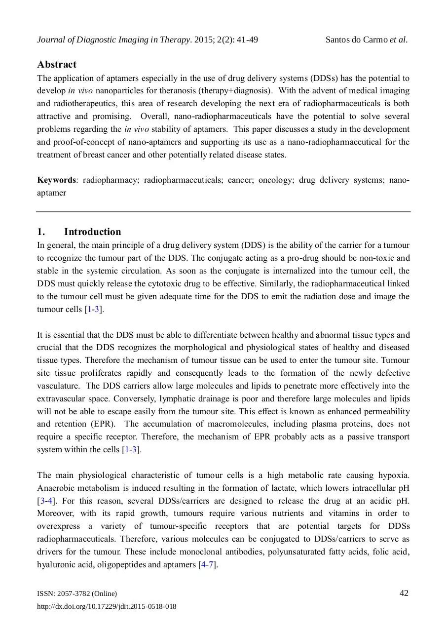
commercial production of designer antibodies. These studies have shown several advantages of using
aptamer technology over antibody therapies and the nano-aptamer demonstrated a size of between 100200 nm and was spherical in shape (Figure 1). This nano-aptamer size is particularly favourable for
medical application because it allows the usage of several imaging modalities such as SPECT and CT
imaging.
Figure 1. Nano-aptamer with a size variation between 100-200 nm.
5.1
Cytotoxicity
To determine the survival of both normal and cancer cells after nano-aptamer treatment, we utilised the
spectrophotometric method, in conjunction with XTT. The graphs below (Figure 2) show the
comparison of the nanoparticle towards various cell types. In the case of MDA-MB 231 and MCF-7, it
was noticed that the nano-aptamer at a dose of 500 μg/mL decreased cell survival to 80%. However,
the aim of this study is to demonstrate that the nano-aptamer is capable of killing tumour cells our data
also supports the migration of the nanoparticle to the particular site of the tumour. These observations
were confirmed by scintigraphic imaging.
ISSN: 2057-3782 (Online)
http://dx.doi.org/10.17229/jdit.2015-0518-018
45
Journal of Diagnostic Imaging in Therapy. 2015; 2(2): 41-49
Santos do Carmo et al.
Figure 2. The effect of aptamer nanoparticles on the survival of A549, MCF-7, MDA-MB 231,
Neuro-2a and SKOV-3 cells. The cells in RPMI/DMEM underwent treatment with various
concentrations of nanoparticles for 72 h. The survival rate was estimated using the MTT assay with
measured values (mean ± SD; n ≥ 3). These were calculated as a percentage of those obtained from the
untreated control (*p < 0.05).
5.2
Biodistribution
Figure 3 indicates the onset of the nanoparticles biodistribution to breast cancer; this is located on the
flank of the animal and invisible to the migration site. Although all the necessary precautions were
taken regarding the welfare of the mice, a number of them eventually died during the imaging process.
This unfortunate limitation was resultant in hampering the imaging process.
ISSN: 2057-3782 (Online)
http://dx.doi.org/10.17229/jdit.2015-0518-018
46
Journal of Diagnostic Imaging in Therapy. 2015; 2(2): 41-49
Santos do Carmo et al.
Figure 3. Scintigraphy of nano-aptamers in the breast cancer animal model.
The methodology to obtain nano-aptamers does appear to be quite attractive. The usage of aptamers as
nanoparticles is promising, as one of the major problems related to aptamers is the rapid efficacy-loss
due to the tumour microenvironment, especially the pH. The aptamers seem not to exist encapsulated
and therefore long-circulating aptamers were present.
6.
Conclusion
In this study, we have demonstrated the application of an aptamer nanoparticle to image breast tumours
in mice. Furthermore, it is important to realize that the encapsulation of the aptamer does not interfere
with the position of the tumour site. In the future, this opens new possibilities for the design and
development of innovative nano-radiopharmaceuticals for diagnostic imaging and treatment plans.
Conflicts of Interest
The authors report no conflicts of interest.
ISSN: 2057-3782 (Online)
http://dx.doi.org/10.17229/jdit.2015-0518-018
47
Journal of Diagnostic Imaging in Therapy. 2015; 2(2): 41-49
Santos do Carmo et al.
References
Key References: 1, 2, 11, 13, 21
[1]
Itakura S, Hama S, Ohgita T, Kogure K. Development of nanoparticles incorporating a novel liposomal membrane
destabilization peptide for efficient release of cargos into cancer cells. PLoS One. 2014; 9(10): e111181.
[CrossRef] [PubMed Abstract]
[2]
Makino A, Kimura S. Solid tumor-targeting theranostic polymer nanoparticle in nuclear medicinal fields.
ScientificWorldJournal. 2014; 2014: 424513.
[CrossRef] [PubMed Abstract]
[3]
Sakurai Y, Kajimoto K, Hatakeyama H, Harashima H. Advances in an active and passive targeting to tumor and
adipose tissues. Expert Opin Drug Deliv. 2015; 12(1): 41-52.
[CrossRef] [PubMed Abstract]
[4]
Kong RM, Ding L, Wang Z, You J, Qu F. A novel aptamer-functionalized MoS2 nanosheet fluorescent biosensor
for sensitive detection of prostate specific antigen. Anal Bioanal Chem. 2015; 407(2): 369-377.
[CrossRef] [PubMed Abstract]
[5]
de Franciscis V. A theranostic "SMART" aptamer for targeted therapy of prostate cancer. Mol Ther. 2014;
22(11):1886-1888.
[CrossRef] [PubMed Abstract]
[6]
Le TT, Adamiak B, Benton DJ, et al. Aptamer-based biosensors for the rapid visual detection of flu viruses. Chem
Commun (Camb). 2014; 50(98): 15533-15536.
[CrossRef] [PubMed Abstract]
[7]
Sá LT, Pessoa C, Meira AS, da Silva MI, Missailidis S, Santos-Oliveira R. Development of nanoaptamers using a
mesoporous silica model labeled with (99m)tc for cancer targeting. Oncology. 2012; 82(4): 213-217.
[Reference Source] [PubMed Abstract]
[8]
Ara MN, Matsuda T, Hyodo M, et al. Construction of an aptamer modified liposomal system targeted to tumour
endothelial cells. Biol Pharm Bull. 2014; 37(11): 1742-1749.
[CrossRef] [PubMed Abstract]
[9]
Manandhar Y, Bahadur KC, Wang W, Uzawa T, Aigaki T, Ito Y. In vitro selection of a peptide aptamer that
changes fluorescence in response to verotoxin. Biotechnol Lett. 2015; 37(3): 619-625.
[CrossRef] [PubMed Abstract]
[10]
Zhou C, Chen T, Wu C, et al. Aptamer CaCO3 nanostructures: a facile, pH-responsive, specific platform for
targeted anticancer theranostics. Chem Asian J. 2015; 10(1): 166-171.
[Reference Source] [PubMed Abstract]
[11]
Sá LT, Simmons S, Missailidis S, da Silva MI, Santos-Oliveira R. Aptamer-based nanoparticles for cancer
targeting. J Drug Target. 2013; 21(5): 427-434.
[Reference Source] [PubMed Abstract]
[12]
Niazi JH, Verma SK, Niazi S, Qureshi A. In vitro HER2 protein-induced affinity dissociation of carbon nanotubewrapped anti-HER2 aptamers for HER2 protein detection. Analyst. 2015; 140(1): 243-249.
[CrossRef] [PubMed Abstract]
[13]
Perkins A, Missailidis S. Radiolabelled aptamers for tumour imaging and therapy. Q J Nucl Med Mol Imaging.
2007; 51(4): 292-296.
[Reference Source] [PubMed Abstract]
ISSN: 2057-3782 (Online)
http://dx.doi.org/10.17229/jdit.2015-0518-018
48
Journal of Diagnostic Imaging in Therapy. 2015; 2(2): 41-49
Santos do Carmo et al.
[14]
Borbas KE, Ferreira CSM, Perkins A, Bruce JI, Missailidis S. Design and synthesis of mono- and multimeric
targeted radiopharmaceuticals based on novel cyclen ligands coupled to anti-MUC1 aptamers for the diagnostic
imaging and targeted radiotherapy of cancer. Bioconjugate Chem. 2007; 18(4): 1205-1212.
[CrossRef]
[15]
Missailidis S, Perkins A. Aptamers as novel radiopharmaceuticals: Their applications and future prospects in
diagnosis and therapy. Cancer Biother Radiopharm. 2007; 22(4): 453-468.
[Reference Source] [PubMed Abstract]
[16]
Ferreira CSM, Cheung MC, Missailidis S, Bisland S, Gariépy J. Phototoxic aptamers selectively enter and kill
epithelial cancer cells. Nucleic Acids Res. 2009; 37(3): 866–876.
[CrossRef] [PubMed Abstract]
[17]
Ferreira CSM, Papamichael K, Guilbault, G, Schwarzacher T, Gariepy J, Missailidis S. DNA aptamers against
MUC1: Design of aptamer-antibody sandwich ELISA for early tumour diagnosis. Anal Bioanal Chem. 2008;
390(4): 1039-1050.
[Reference Source] [PubMed Abstract]
[18]
DaPieve C, Perkins AC, Missailidis S. Anti-MUC1 aptamers: radiolabelling with
MCF-7 tumour-bearing mice. Nuclear Med Biol. 2009; 36: 703-710.
[Reference Source] [PubMed Abstract]
[19]
Leite Diniz C, Da Pieve C, Perkins A, et al. 1.227 Pharmacokinetic and biodistribution studies of anti MUC1
PEGylated aptamers with potential in the targeted radiotherapy of breast cancer. EJC Supplements. 2010; 8(5): 5960.
[CrossRef]
[20]
Patricio BFC, Weissmuller G, Santos-Oliveira R. Development of the 153-SM-EDTMP nanoradiopharmaceutical.
J Label Compd Radiopharm. 2011; 54: S558.
[Reference Source]
[21]
Patricio BFC, Albernaz MS, Sarcinelli MA, de Carvalho SM, Santos-Oliveira R, Weissmüller G. Development of
novel nanoparticle for bone cancer. J Biomed Nanotechnol. 2014; 10(7): 1242-1248.
[CrossRef] [PubMed Abstract]
[22]
Pires J, Patrico BFC, Albernaz MS, et al. Preparation of biodegradable poly(L-Lactide) (PLA) nanoparticles
containing DMSA (dimercaptosuccinic acid) as novel radiopharmaceutical. Advanced Science Letters. 2012;
10(1): 143-145.
[CrossRef]
99m
Tc and biodistribution in
Citation: Santos do Carmo F, Rocha Pinto S, Maria Camões Orlando M, et al. Nano-Aptamer for
Breast Cancer Imaging: Initial Considerations. Journal of Diagnostic Imaging in Therapy. 2015; 2(2):
41-49.
DOI: http://dx.doi.org/10.17229/jdit.2015-0518-018
Copyright: © 2015 Santos do Carmo F, et al. This is an open-access article distributed under the terms
of the Creative Commons Attribution License, which permits unrestricted use, distribution, and
reproduction in any medium, provided the original author and source are cited.
Received: 07 May 2015 | Revised: 15 May 2015 | Accepted: 16 May 2015
Published Online 18 May 2015 http://www.openmedscience.com
ISSN: 2057-3782 (Online)
http://dx.doi.org/10.17229/jdit.2015-0518-018
49
Download JDIT-2015-0518-018
JDIT-2015-0518-018.pdf (PDF, 603.16 KB)
Download PDF
Share this file on social networks
Link to this page
Permanent link
Use the permanent link to the download page to share your document on Facebook, Twitter, LinkedIn, or directly with a contact by e-Mail, Messenger, Whatsapp, Line..
Short link
Use the short link to share your document on Twitter or by text message (SMS)
HTML Code
Copy the following HTML code to share your document on a Website or Blog
QR Code to this page

This file has been shared publicly by a user of PDF Archive.
Document ID: 0000603708.