Biomaterials 31 (2010) 3762–3771
Contents lists available at ScienceDirect
Biomaterials
journal homepage: www.elsevier.com/locate/biomaterials
Neuronal adhesion and differentiation driven by nanoscale surface
free-energy gradients
Guillaume Lamour a, b, Ali Eftekhari-Bafrooei b, Eric Borguet b, Sylvie Soue`s c, Ahmed Hamraoui a, d, *
a
Neuro-Physique Cellulaire, Universite´ Paris Descartes, UFR Biome´dicale, 45 Rue des Saints-Pe`res, 75006 Paris, France
Department of Chemistry, Temple University, Philadelphia, Pennsylvania, PA 19122, USA
c
Re´gulation de la Transcription et Maladies Ge´ne´tiques, CNRS UPR2228, Universite´ Paris Descartes, UFR Biome´dicale, 45 Rue des Saints-Pe`res, 75006 Paris, France
d
Service de Physique et Chimie des Surfaces et Interfaces, CEA Saclay, 91191 Gif-sur-Yvette, France
b
a r t i c l e i n f o
a b s t r a c t
Article history:
Received 16 December 2009
Accepted 15 January 2010
Available online 10 February 2010
Recent results indicate that, in addition to chemical, spatial and mechanical cues, substrate physical cues
such as gradients in surface energy may also impact cell functions, such as neuronal differentiation of
PC12 cells. However, it remains to be determined what surface effect is the most critical in triggering
PC12 cell differentiation. Here we show that, beyond continuously probing the surface energy landscape
of their environment, PC12 cells are highly sensitive to nanoscale chemical heterogeneities. Selfassembled monolayers of alkylsiloxanes on glass were used as a culture substrate. By changing the
structure, ordering and chemical nature of the monolayer, the surface energy distribution is altered.
While both well-ordered CH3 terminated substrates and bare glass (OH terminated) substrates did not
favor PC12 cell adhesion, PC12 cells seeded on highly disordered CH3/OH substrates underwent
enhanced adhesion and prompt neuritogenesis by 48 h of culture, without nerve growth factor treatment. These data illustrate that surface free-energy gradients, generated by nanoscale chemical
heterogeneities, are critical to biological processes such as nerve regeneration on biomaterials.
Ó 2010 Elsevier Ltd. All rights reserved.
Keywords:
PC12 cells
Neuronal differentiation
Cell adhesion
Self-assembled monolayers (SAMs)
Sum-frequency generation (SFG)
Surface energy
1. Introduction
Neuronal differentiation is critical to nervous tissue regeneration after injury, and adhesion on a substrate is critical for neurite
extension [1–3]. The initiation and guidance of a neurite rely on
extra-cellular signals, such as substrate energy of adhesion (e.g.,
surface energy, or surface tension) [4], especially local gradients [5].
Hence, it is of great interest to unveil the substrates characteristics
that are effectively sensed by the growth cone, and translated into
neuritis extension as a response to these physical cues. The ability
to spatially control the distribution of the energy of adhesion is of
particular interest in many biomedical and tissue-engineering
applications.
The interactions of cells, especially neurons, with nanoscale
topography [6–9], and with surface chemistry [10–12], were
reported to be important parameters in controlling cell function.
Another parameter, substrate compliance, influences both neuritogenesis [13–16] and neurite branching rate [17]. Thus, a combination of spatial, chemical and mechanical inputs, together with
the genetic program of the cell, has been recently proposed to
* Corresponding author. Fax: þ33 (0)14 286 2085.
E-mail address: ahmed.hamraoui@parisdescartes.fr (A. Hamraoui).
0142-9612/$ – see front matter Ó 2010 Elsevier Ltd. All rights reserved.
doi:10.1016/j.biomaterials.2010.01.099
control the shape and functions of cells, as well as of tissues [18].
However the exact role of the surface tension and its spatial variation is still unclear and a systematic study may lead to a better
understanding of the surface adhesion parameters that drive
neuritogenesis.
Self-assembled monolayers (SAMs) are surface-active materials
with many potential applications in biotechnology [19,20]. SAMs of
alkylsiloxanes on glass can exhibit a wide range of properties,
including the chemical nature, the surface roughness, and the
organization of surface-exposed terminal groups. These combined
properties generate, at the nanoscale level, distinct surface energy
distributions, and at larger scale, macroscopic surface characteristics such as wettability. SAMs of alkylsiloxanes on glass [21,22] or
on titanium [23] have been shown to be suitable substrates for
controlling cell adhesion, and in particular for controlling neuronal
cell differentiation [10,21,23].
PC12 cells, though not primary neuronal cells, express the
transmembrane TrkA and p75 receptors to nerve growth factor (NGF)
[24,25], and differentiate into a neuronal phenotype when challenged by appropriate NGF concentrations [26]. This ability makes
them a well-defined model to study neuronal differentiation mechanisms, and thus axonal regeneration. Several key inducers of PC12
cell neuronal differentiation in NGF-free medium have been identified: PC12 cell neuritogenesis is observed on soft substrates
�
G. Lamour et al. / Biomaterials 31 (2010) 3762–3771
3763
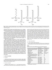
Fig. 1. Schematics of methyl-terminated molecules used to modify glass surfaces. Molecules were grafted onto clean glass surfaces by chemisorption from the liquid phase. HTMS,
OTMS and OTS cross-link during SAMs formation, contrary to ODMS or ODS, that can bind glass surfaces through only one bond, following hydrolysis of their unique OCH3 or
chlorine leaving group.
composed of extra-cellular matrix (ECM) proteins such as collagen,
fibronectin and laminin [27], or of ECM derived from astrocytes [28].
In our previous study [5], we demonstrated the differentiation
ability of PC12 cells in NGF-free medium when seeded on solid glass
substrates covered with NH2-terminated alkylsiloxane SAMs. These
surfaces contained a nanoscale mixture of hydroxyl and amine
groups which provided local gradients in surface energy. However,
we did not determine whether the trigger of PC12 cell differentiation was the surface nanoroughness, the surface concentration in
terminal amines, the alternation of OH and NH2 groups, or
a combination of these factors.
Here we examined the influence of these potential triggers,
again by tailoring various silanes on glass [29], which provide cell
culture substrates. We used methyl-terminated silanes that offer
two main advantages compared to aminosilanes. First, because of
the smaller reactivity of CH3 compared to NH2 groups, the control
over adsorption process is easier. It is noted that the highly reactive
leaving groups of silanes molecules (chlorine and methoxy) react
with the silanol groups and/or adsorbed water on the glass surface
and are present on the side of the molecule close to the glass
surface, away from CH3 terminal groups (Fig. 1). Second, vibrational
spectroscopy which is used for the surface characterization is more
reliable in the CH stretching region (2800–3000 cm�1) than in the
NH stretching region (3100–3500 cm�1) where the broad OH
vibrational spectrum of adsorbed water smears out the NH peaks.
Furthermore, CH groups do not provide polar component to the
surface free energy (SFE), thus facilitating the SFE calculations and
SFE analysis.
2. Materials and methods
2.1. Chemicals
Chemicals were obtained from Acros Organics (Geel, Belgium), Sigma–Aldrich
(St. Quentin Fallavier, France), ABCR (Karlsruhe, Germany), Fisher Scientific (Illkirch,
France) and Carlo Erba Reagents (Val de Reuil, France). The sources and purity of the
chemicals used are summarized in Table 1.
2.2. Substrates preparation
Modified glass slides (SuperFrostÒ, 25 � 75 � 1 mm3, Menzel-Glaser, Braunschweig, Germany) were used for optical studies, and modified glass coverslips (30-mm
diameter and 100-mm thick, Menzel-Glaser) were used for cell culture experiments.
Prior to use, glassware was cleaned by immersion in piranha solution (3:1 (v/v)
sulfuric acid:40% hydrogen peroxide), then thoroughly rinsed with deionized water
and dried under a nitrogen stream (caution: piranha solution is extremely corrosive
and can react violently with organic compounds. Appropriate safety precautions
including gloves and face shield should be used when handling.). Glass coverslips were
cleaned by immersion in ultrasonic bath of chloroform for 20 min prior to immersion in piranha solution. For the self-assembly, the cleaned glass substrates were
immersed into solutions (Table 2) of the desired alkylsilanes (Fig. 1). The chemically
modified substrates were then rinsed with the neat solvent. Prior to cell culture, the
substrates were dried under a laminar flow hood and prior to surface characterization, the substrates were dried under a nitrogen stream. All treatments were
carried out at room temperature and in ambient atmosphere (relative
humidity z 50%).
2.3. Surface characterization
2.3.1. Fourier transform infrared spectroscopy
Fourier transform infrared spectroscopy (FTIR) spectra were measured in the
transmission geometry at a normal incidence angle using a Bruker Optics TENSOR 27
Table 1
Chemicals used for surface modification and contact angle measurements.
Deionized water (Elga UHQ PS MK3)
Hexanes (HX) (mixture of isomers)
Methanol (MET)
Acetic acid (AA)
Sulfuric acid
Hydrogen peroxide
Chloroform (CF)
n-Hexyltrimethoxysilane (HTMS)
n-Octadecyltrimethoxysilane (OTMS)
Octadecyldimethylmethoxysilane
(ODMS)
n-Octadecyltrichlorosilane (OTS)
Octadecyldimethylchlorosilane (ODS)
Glycerol (GL)
Formamide (FA)
n-Hexadecane (HD)
Tetradecane (TD)
n-Dodecane
n-Undecane
n-Octane
a
Water content is <0.01% (v/v).
Manufacturer
Purity (%)
Veolia Water Systems
Sigma–Aldrich
Carlo Erba Reagents
Carlo Erba Reagents
Sigma–Aldrich
Sigma–Aldrich
Carlo Erba Reagents
ABCR
Acros Organics
ABCR
(r ¼ 18.2 MU.cm)
�98.5 (ACS)a
>99.9 (HPLC)a
99.9 (RPE)
95–97
40 (m/v in H2O)
>99.8 (ACS)
97
95
95
Acros Organics
Sigma–Aldrich
Acros Organics
Fisher Scientific
Acros Organics
Sigma–Aldrich
Fisher Scientific
Acros Organics
Acros Organics
95
95
�99
�99.5
99
�99
>98
99
95
�
3764
G. Lamour et al. / Biomaterials 31 (2010) 3762–3771
Table 2
Description of the chemical processes used to modify glassware. The abbreviations
in capital letters refer to the chemicals displayed in Table 1.
Substrate
Solution
Adsorption
time
Rinsing
solvent(s)
Note(s)
ots
ods
otms
otmsx
odms1
odms2
htmsM1
0.1% OTS þ 20% CF þ 80% HD
0.1% ODS þ 20% CF þ 80% HD
1% OTMS þ 99% HX
1% OTMS þ 99% HX
1% ODMS þ 99% HX
1% ODMS þ 99% HX
2% HTMS þ 94% MET þ 4%
H2O þ 1 mM AA
2% HTMS þ 94% MET þ 4%
H2O þ 1 mM AA
2% HTMS þ 94% MET þ 4%
H2O þ 1 mM AA
1% HTMS þ 99% HX
1% HTMS þ 99% HX
15 min
15 min
4h
4h
w10 h
>24 h
w16 h
CF and MET
CF and MET
HX and MET
HX and MET
HX and MET
HX and MET
MET
a
w24 h
MET
>72 h
MET
4h
4h
HX and MET
HX and MET
htmsM2
htmsM3
htmsH
htmsHx
a,b
a
a
a
a
Solution was slowly agitated with a rotating magnet all along the reaction.
ODS was slightly heated until it reached a liquid phase (at 28–30 � C), just before
being added to the solvent solution.
Table 3
Values of the surface tension (g, mN m�1) of some test liquids at 20 � C (adapted from
Ref. [33]). gd and gp are respectively the dispersive and the polar components of the
surface tension.
Liquid
g
gd
gp
Water
Glycerol
Formamide
n-Hexadecane
Tetradecane
72.8
64
58
27.47
26.56
21.8
34
39
27.47
26.56
51
30
19
w0
w0
2.3.5. AFM imaging
Each substrate was analyzed using a BioscopeÔ AFM (Digital Instruments/
Veeco) in air using tapping mode (RTESP tip cantilever, spring constant: 40 N m�1).
The root-mean-square (rms) roughness of the surfaces was evaluated for regions of
w1 mm � 1 mm, by AFM software Nanoscope (Veeco). The line scanning frequency
was w0.5 Hz (256 scan lines � 512 pixels). The images were flattened using Nanoscope software before the rms was evaluated.
b
2.4. PC12 cell manipulation
Unless otherwise specified, the biological products in this section were
purchased from Invitrogen (Fisher Bioblock Scientific, Illkirch, France).
spectrometer equipped with a DTGS detector. FTIR spectra were background corrected by subtraction of a spectrum of the clean, bare substrate (e.g., SAM-free), and
recorded by integrating 200 scans with a resolution of 4 cm�1.
2.3.2. Vibrational sum-frequency generation
Sum-frequency generation (SFG) is a surface vibrational spectroscopy based on
a second-order non-linear optical process in which two laser beams with frequencies u1 and u2 overlap on the surface and generate a coherent response whose
frequency is the sum of two incident laser (uSFG ¼ u1 þ u2). In the electric dipole
approximation, second-order non-linear optical processes, including SFG, do not
take place in media with inversion symmetry but do occur at interfaces as the
inversion symmetry is necessarily broken there. Therefore, by selecting one of the
incident beams to be in the IR region (u1 ¼ uIR) and the other in the visible region
(u2 ¼ uvis.), SFG can be used as a powerful spectroscopic technique to measure
surface vibrational spectra [30]. A detailed description of our SFG set up can be found
elsewhere [31]. Briefly, IR and visible pulses with energies of 15 and 2 mJ, incident at
the surface with the angles of 72� and 65� respectively, were focused to beam waists
of 250 and 200 mm, respectively. The SFG signal was detected with a CCD (Princeton
Instrument) coupled with a spectrograph (300i, Acton Research Corp.). The polarization of visible and SFG were controlled by a combination of polarizers and halfwave plates. In the experiments presented here, the polarization combination of
SFG, visible, and IR were either S,S,P or P,P,P.
2.4.1. Cell culture
PC12 cells (ATCC, CRL 1721) were maintained in Dulbecco’s Modified Eagle
Medium containing horse serum (5%), fetal calf serum (5%, HyClone), non-essential
amino acids (1%) and antibiotics (1%). In the experiments, PC12 cells (passage
numbers 7–17) were seeded onto modified glass coverslips, that had been sterilized
by immersion in a solution of 70% methanol and 30% H2O for 15 min. Cells were
seeded in a small volume of the culture medium (V ¼ 335 mL), in order to trap PC12
cells on the top of the modified substrates. The cell density at the time of seeding
was w104 cm�2. Experiments never exceeded 48 h. No further addition of culture
medium was made and, in particular, no NGF was added to the culture medium.
2.4.2. Quantification of neuritogenesis
The propensity of PC12 cells to initiate neurites was evaluated on each substrate.
At least 10 pictures (S ¼ 0.182 mm2/picture) of the cells cultured on each substrate
were taken with a camera mounted on a phase-contrast microscope (Nikon Eclipse
TS100), using a �20 objective. In order to avoid confusing neurites with cell soma
protrusions or filopodia, only neurites with length greater than 25 mm were counted.
This threshold length corresponds to approximately the diameter of a PC12 cell
soma multiplied by 1.5.
2.3.4. Determination of surface free energy
The surface free energies (SFE) of samples were calculated using the Owens–
Wendt theoretical model [32]. This model gives the long-range dispersion (Lifshitz–
Van der Waals; gd) and the short-range polar (hydrogen bonding; gp) components of
SFE according to the following equation:
2.4.3. Immunofluorescence
PC12 cells were cultured on each substrate, as described above. After 48 h of
culture, cells were fixed with 3.7% formaldehyde in PBS for 15 min and then permeabilized with 0.1% Triton X-100 in PBS/0.1% bovine serum albumin (BSA) for
20 min. All washes, blocking steps, and antibody dilutions were performed using
0.1% BSA, 0.01% TritonÒ X-100 in PBS. After cell fixation and permeabilization, the
primary antibody anti-MAP1B (Sigma–Aldrich; diluted 1:800) was incubated overnight at 4 � C. A secondary Cy3-conjugated antibody (Jackson ImmunoResearch,
Cambridgeshire, UK; diluted 1:400) was incubated for 2 h at room temperature. DNA
was stained with 4-6-diamidino-2-phenylindole (DAPI) at 0.5 g/mL for 15 min.
F-actin was stained with phalloidin coupled to Alexa Fluor 488 (Molecular Probes) at
5 units/mL for 30 min. Finally cells were extensively washed in PBS and mounted in
a Fluoromount GÔ solution (Southern Biotech). Cell observation was done with
a Nikon Eclipse E600 epifluorescence microscope coupled to a high resolution colour
camera (Nikon DXM 1200), using �10 and �50 objectives. No threshold processing
was applied to the images.
�
�1=2 d �1=2
�1=2 p �1=2
gL
gL
þ 2 gps
WSL ¼ 1 þ cos q gL ¼ 2 gds
3. Results and discussion
2.3.3. Contact angle measurements
Contact angles (q) were measured as described in Ref. [5]. Briefly, an image of
the profile of a drop on a solid surface was recorded using a CCD video camera
(Sony DXC-101P). The image was then processed with ImageJ software (Wayne
Rasband, National Institutes of Health, Bethesda, MD) using the Contact Angle
plug-in (Marco Brugnara, University of Trento, Trento, Italy), which calculates the
contact angle value from the profile of the drop. At least 3 drops per sample were
analyzed.
(1)
where gs is the SFE of the surface, gL is the SFE of the liquid and WSL is the solid–
liquid interface energy. Two liquids were used as probes for SFE calculations:
n-hexadecane, and H2O. For a liquid, the overall surface tension (gL) is a combination
of dispersive and polar components, whose values are indicated in Table 3 (adapted
from Ref. [33]). The contact angles of n-hexadecane and H2O were reported in Eq. (1)
for each solid substrate, then sample SFE components were calculated.
The critical surface tension gc was calculated using the Fox–Zisman
approximation. In this work, it can be understood as a first-order approximation
of the Good–Girifalco equation [35], for a surface tension of the liquid gL (gL � gc)
close to the gc of the solid. Using a linear regression analysis, Zisman plots,
cos q ¼ f(g), were traced for each substrate by fitting the data obtained with the
test liquids. gc values were read where the line fits intersect cos q ¼ 1, as
described by Zisman [34].
Molecules (noted in capital letters) of various chain lengths
and chemical nature (Fig. 1) were chosen to generate distinct
surfaces (noted in small letters), according to their mechanism of
adsorption and monolayer formation on glass. The resulting
substrates were divided into three different classes (Fig. 2) with
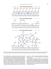
regard to nanoscale surface organization and hence to distribution of surface energy. Class 1 SAMs are well-ordered with an alltrans conformation of the alkyl chains. Class 2 SAMs, while
eliminating multilayer formation due to the impossibility of the
monomers undergoing cross-linking, are disordered. Moreover,
�
G. Lamour et al. / Biomaterials 31 (2010) 3762–3771
3765
Fig. 2. Sketches representing three distinct organizations of SAMs used as substrates for PC12 cell culture. Class 1 SAMs are well-ordered with an all-trans conformation of alkyl chains.
Class 2 SAMs are disordered but limited to monolayer formation. Class 3 SAMs are highly disordered, with possible multilayer formation and higher chemical heterogeneity.
class 2 SAMs likely are accompanied by a significant density of
substrate silanol groups (Si–OH), compared to class 1 SAMs. This
is a consequence of class 2 monolayer adsorption being incomplete by nature. Class 3 SAMs are the result of chaotic polymerization of a trialkoxysilane. The chemisorption of siloxanes is
believed to require activation of the siloxane whereby the leaving
group (e.g., OCH3 in HTMS) is replaced by OH from water. When
the solvent solution contains more than the trace amount of
water necessary for adsorption reaction to occur, methoxy groups
are quickly hydrolyzed before chemical adsorption on the silica
surface. As a result, the molecules polymerize through their
silanol groups. It is the resultant polymer that chemically binds
the silica surface.
In view of intrinsic properties of both molecules and solvent
solutions (Table 2) it was expected that class 1 SAMs would be
generated by OTS and OTMS molecules, that class 2 SAMs would be
the result of ODS and ODMS adsorption, and that HTMS would
generate either class 1 or class 3 SAMs according to the solvent
solution used. The particular properties of each class of substrates
are described here after.
�
3766
G. Lamour et al. / Biomaterials 31 (2010) 3762–3771
discrepancy could result from monomers cross-linking, probable
with HTMS, but impossible with ODMS. The binding of HTMS
monomers to each other laterally would increase the number of
adsorbed molecules per area. Surprisingly, the intensity of the CH3
peak of htmsH substrate was even greater than that of the ots
substrate. AFM experiments revealed that the rms roughness of
htmsH substrate is remarkably higher (w1.4 nm) than that of the
others substrates (w0.3 nm) which are supposed to display one
single organic monolayer on the bare glass surface. These results
suggest that the htmsH modified surface is partially over covered by
additional layers of HTMS molecules, possibly physisorbed on it. To
determine whether the glass-bound layer of HTMS, and of the other
molecules was coherently organized, a non-linear spectroscopic
analysis was performed.
3.2. Qualitative analysis of surface-exposed CH groups
organization by SFG
Fig. 3. FTIR spectra in the CH region of some substrates. FTIR spectra were background
corrected by subtraction of a spectrum of the clean glass substrate (e.g., SAM-free).
Spectra are offset for clarity. Peaks at w2850 cm�1, w2920 cm�1, and w2955 cm�1 are
respectively assigned to CH2-ss, CH2-as, and CH3-as. Surface density of glass-bound
alkylsiloxanes is reflected by the intensity of peaks, that depends on molecules alkyl
chain length (for CH2 peaks only) and on molecules organization inside the monolayers. This latter aspect is developed in the SFG spectra (Fig. 4) of the same substrates.
The dashed line at w2018 cm�1 represents close packed long chain SAM obtained for
the ots substrate. Values in parentheses indicate the water contact angle on each
substrate.
3.1. Quantitative analysis of substrates by FTIR spectroscopy
Modified substrates were probed by FTIR in the CH stretching
region (Fig. 3). The ‘‘ideal’’ and well-studied monolayer made of OTS
molecules is known to lead to a full coverage of the bare glass surface
[36]. As expected, its FTIR spectrum in the CH region displayed the
most intense peaks among all other SAMs studied. The CH peak
intensity of odms substrates was lower than those of ots substrates,
although the alkyl chain length is identical (Fig. 1). This indicates
that odms1 and odms2 substrates have fewer molecules grafted per
unit area. The higher intensity of CH2 symmetric (CH2-ss) and
asymmetric (CH2-as) stretches, in odms2 compared to odms1
substrate, can result from a higher quantity of adsorbed material, as
supported by the higher contact angle value. In contrast to these
surfaces, the htmsM3 substrates showed almost no visible CH peak,
suggesting that the quantity of material grafted is under the detection limit of the FTIR spectrometer. Nevertheless, the presence of
a non-negligible amount of CH groups at htmsM3 surface was suggested by the relatively high contact angle measured for water (56� )
while a bare glass surface would undergo complete wetting
(e.g., qH2O z 0� ).
The intensity of the CH peaks of htmsH substrates was similar to
that of odms substrates, although the HTMS molecule contains
fewer CH2 groups than ODMS (5 versus 17). This apparent
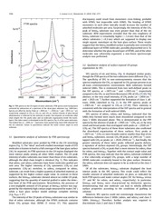
SFG spectra of ots and htmsH (Fig. 4) displayed similar peaks,
though the FTIR spectra of the two substrates were different (Fig. 3).
The specificity of SFG to non-centrosymmetry ensures that the
spectra emphasize the CH3, as opposed to CH2 groups present in an
all-trans centrosymmetric environment. Both substrates formed
ordered SAMs. This is evidenced from two well-defined peaks in
the SSP spectra, at w2875 cm�1 and w2955 cm�1, respectively
assigned to the CH3-ss and Fermi Resonance (FR) of CH3 [37,38]. The
absence, or the very small intensity, of CH2 peaks (at w2850 cm�1
and w2925 cm�1) further supported the formation of class 1, alltrans, SAMs (sketched in Fig. 2). In the PPP spectra, peaks at
w2965 cm�1 are assigned to CH3-as [37,38]. Their intensity is
consistent with the interpretation of the SSP spectra and in general
with the formation of well-ordered SAMs.
SFG spectra of odms1, odms2 and htmsM3 indicated that the
SAMs they formed were much more disordered compared to the
class 1 SAMs discussed above. This is demonstrated in the PPP
spectra by the absence of peak at w2965 cm�1 (CH3-as), or by very
small and broad peaks that overlapped with peaks at w2930 cm�1
(CH2-as). The SSP spectra of these three substrates also confirmed
the disordered organization of these surfaces. First, peaks at
w2875 cm�1 (CH3-ss) were broader and/or smaller than that of ots
and htmsH substrates, second, CH3-FR peaks (at 2955 cm�1) overlapped with CH2-as peaks (at w2920–2930 cm�1). And finally, the
greater intensity of these latter peaks reflected gauche defects,
a signature of surface-exposed CH2 groups. Interestingly, the SSP
spectra featured a CH3-ss peak that is more intense for odms2 than
for odms1 and htmsM3. Together with the contact angle value and
FTIR spectra, this may indicate that the odms2 substrate exhibited
a few coherently arranged CH3 groups, with a large number of
ODMS molecules covalently bound to the glass surface. However,
the odms2 substrate does not appear to be as ordered as the class 1
SAMs.
Of all the substrates, htmsM3 substrate displayed the least
intense peaks in the SFG spectra. This result could reflect the
smaller amount of adsorbed molecules on glass, as indicated by
both FTIR spectra and water contact angles (56� for htmsM3 versus
77� and 96� for odms1 and odms2, respectively). In particular,
htmsM3 and htmsH were characterised by distinctive spectra,
demonstrating that one molecule can lead to wholly different
surface properties according to the conditions of grafting in
solution.
However, SFG spectra could not convincingly characterize the
differences between class 2 SAMs (odms1 and odms2) and class 3
SAMs (htmsM3). Therefore, further analysis was required to
discriminate class 2 and class 3 SAMs.
�
G. Lamour et al. / Biomaterials 31 (2010) 3762–3771
3767
Fig. 4. SFG spectra of SAMs used as model culture substrates and PC12 cell adhesion and differentiation, without NGF treatment, on substrates 48 h after seeding. Top graphs feature
SFG traces in the CH region for the substrates also analyzed by FTIR in Fig. 3. Spectra are offset for clarity. The polarization combination for SFG, visible and IR is S,S,P for left top
graph, and P,P,P for right top graph. Clear surface organization (e.g., intense CH3-group peaks and absence of CH2 peaks) arises in class 1 substrates, compared to the others. Values in
parentheses indicate the water contact angle on each substrate. PC12 cells have three distinct fates according to substrate class: no adhesion on class 1 SAM, relatively poor adhesion
and neurite outgrowth on class 2 SAM, and good adhesion preceding neuronal differentiation on class 3 SAM. Observations were made 48 h after seeding with a contrast phase
microscope.
3.3. SFE determination by contact angle measurements
Data obtained for contact angles, SFE, and roughness analysis,
are recapitulated in Table 4. The overall surface tension gs of class 1
substrates ots and htmsH was mostly composed of the dispersive
component gd (Fig. 5), the polar contribution gp being close to zero.
It suggests that very few OH groups were exposed, considering that
gp is not generated by CH3 groups. This result supports the close
arrangement of monomers and its corollary: the complete coverage
of the glass substrate. A similar result was obtained for the otms
substrate, whose SFG spectra resemble those of class 1 SAMs (data
not shown for clarity).
As expected, class 2 substrates odms1, and ods (whose SFG
spectra were similar to those of odms substratesddata not shown),
displayed a more intense gp compared to class 1 substrates. This
suggests the presence of surface-exposed OH groups, in agreement
with the presence of disorganized alkyl chains and low intensity of
FTIR peaks. The surface tension of an almost perfectly homogeneous CH3 substrate is given by the gs of the ots substrate
(w21.6 mN m�1), close to the gc of the same substrate
(w19.9 mN m�1) obtained by a Zisman plot (Fig. 6). For a clean, bare
glass substrate, estimations vary from 150 to 300 mN m�1 [39,40].
Consequently, it is of considerable importance to remark that,
though the added contribution of gp to gs for class 2 substrates is
�
3768
G. Lamour et al. / Biomaterials 31 (2010) 3762–3771
Table 4
Data collected from contact angle measurements and AFM. Contact angles (q) were measured using water, glycerol (GL), formamide (FA), tetradecane (TD), and n-hexadecane
(HD) as test liquids. Uncertainty on a measured contact angle was statistically estimated to less than 1�. Dq is the difference between advancing and receding contact angles of
water. The critical surface tension (gc, �1.5 mN m�1) was determined from Zisman plots, displayed in Fig. 6, using contact angles of all test liquids. The polar ðgps Þ and dispersive
ðgds Þ components of the overall surface tension (gs, �1.0 mN m�1) of the solid substrates were determined from the water and n-hexadecane contact angles. Values of the rootmean-square roughness (�0.1 nm) are the mean of three independent measurements.
qH2O
qGL
qFA
qHD
qTD
gc
gds
gps
gs
rms
Dq
deg
deg
deg
deg
deg
mN m�1
mN m�1
mN m�1
mN m�1
nm
deg
ots
ods
otms
odms1
odms2
htmsM1
htmsM3
htmsH
htmsHx
110
99
90
40
35
19.9
21.5
0.1
21.6
0.3
12
82
77
68
31
7
23
23.7
6.9
30.6
0.3
17
105
92
89
29
15
23.7
24.2
0.3
24.5
0.3
12
77
68
58
13
–
26.7
26.8
8.2
35
0.3
15
96
81
75
24
–
24
25.2
1.6
26.8
0.3
11
38
34
31
6
–
26.5
27.3
32.6
59.9
0.9
32
56
51
45
11
–
25.7
27
20.9
47.9
0.3
22
104
91
83
34
26
22.7
23.1
0.6
23.6
1.4
30
98
87
83
21
12
24.9
25.8
1.2
26.9
0.4
12
relatively small (1.6 mN m�1 � gp � 8.2 mN m�1), locally the
surface energy gradients can reach much higher values
(20 mN m�1 � gc � 150 mN m�1).
Class 3 substrates, e.g., htmsM1 and htmsM3, displayed higher gp
(�20.9 mN m�1) than both class 1 (�0.6 mN m�1) and class 2
substrates (�8.2 mN m�1). The smaller gp of htmsM3
(w20.9 mN m�1) compared to that of htmsM1 (w32.6 mN m�1)
indicates that more HTMS molecules adsorbed on glass in htmsM3.
As htmsM3 was very smooth (rms z 0.3 nm), it appears that HTMS
may have bound to the majority of substrate silanol sites still available for adsorption. Moreover, qH2O could not be further increased by
lengthening the time of adsorption in the methanol/water solution,
supporting the conclusion of HTMS optimal adsorption in these
conditions. Nevertheless, the quantity of adsorbed HTMS was relatively small, as evidenced by the absence of peaks in the htmsM3 FTIR
spectrum (Fig. 3). Therefore the greater part of the gd contribution to
the gs of htmsM3 was provided by OH groups. As a result htmsM
substrates exhibited a chemical pattern that was mostly glass-like,
with a heterogeneous distribution of CH3 groups. Conversely, class 2
SAMs mostly exhibited CH3 groups, together with more (odms1 and
ods) or less (odms2) scattered OH groups.
odms2 and htmsHx are particular substrates. odms2 substrate
concentrated more molecules than odms1, as evidenced by the FTIR
spectra, and its gs components resembled those of class 1
substrates, whose gp was close to zero. However, the gp of odms2
Fig. 5. SFE components gd and gp of solid substrates. gd and gp were calculated
through the measurements of water and n-hexadecane contact angles, displayed in
Table 4, using the Owens–Wendt theoretical model. Notes indicate PC12 cell fate on
substrates 48 h after seeding: either the cells did not adhere (*), or they adhered,
regrouped in clusters, and initiate few neurites (:), or the adhesion was enhanced and
the cells generated many neurites (#).
(w1.6 mN m�1) was higher than the gp of class 1 substrates
(gp < 0.6 mN m�1). Moreover, the SFG spectra evidenced the
disordered organization of CH groups in odms2 substrate. Therefore
odms2 cannot be considered as sharing properties of class 1
substrates. HtmsHx substrate was made by agitating the solution in
order to prevent HTMS molecules from over covering the substrate
(rms z 0.3 nm for htmsHx versus w1.4 nm for htmsH). However,
this process also apparently slowed the reaction kinetics, resulting
in an incomplete monolayer. This is evidenced by qH2O on htmsHx
(98� ), smaller than qH2O on htmsH (104� ). This suggests that the
htmsHx substrate exhibited some OH groups that contributed to
a small gp (w1.2 mN m�1), and though originally forming a class 1
SAM, it did not share the same surface energy distribution. The SFG
spectra of the otmsx substrate featured similar traces than those of
class 2 substrates (data not shown for clarity), and qH2O was also
smaller on otmsx (100� ) than on otms (105� ). Consequently, htmsHx
and otmsx substrates shared the properties of class 2 substrates.
The values of gc, determined by Zisman plots (Fig. 6 and Table 4),
were in agreement with previous results. gc can be assimilated to
gd of solid substrates [41], and the gap between gc and gd never
exceeded 1.6 mN m�1 (Table 4). In addition gc was always smaller in
‘‘ordered’’ substrates than in ‘‘disordered’’ substrates made of
similar molecules. For instance, following relations were obtained:
gc (ots) < gc (ods), gc (otms) < gc (odms2) < gc (odms1), and gc
(htmsH) < gc (htmsHx) < gc (htmsM3) < gc (htmsM1). More generally,
gc values were in the same range (19.9 mN m�1 < gc < 26.7
mN m�1), thus supporting the idea that substrates are rather
comparable in terms of nanoscale SFE distribution than in terms of
overall surface tension.
The difference between advancing and receding contact angles
of water (noted Dq) is a good control to evaluate physical roughness
and/or chemical heterogeneity of surfaces [42–44]. Dq values were
in good agreement with both rms roughness and SFE distribution.
Among smooth substrates whose rms roughness was w0.3 nm,
those generating the smallest gp also generated the smallest Dq
(Table 4). On ods and odms1 substrates, whose gp was higher than
that of ots, otms, odms2, htmsH and htmsHx substrates (Fig. 5 and
Table 4), Dq was higher as well (w15–17� versus w12� ). It was
further higher on htmsM3 (Dq z 22� ) whose surface tension is
locally more heterogeneous. Finally, the largest values (Dq z 30� )
were obtained for substrates exhibiting high roughness: htmsM1
(rms z 0.9 nm) and htmsH (rms z 1.4 nm).
3.4. PC12 cell adhesion and differentiation on modified substrates
When seeded on a clean, bare glass substrate, PC12 cells poorly
adhered and tend to detach by 48 h. On well-ordered SAMs, such
�
G. Lamour et al. / Biomaterials 31 (2010) 3762–3771
3769
Fig. 6. Zisman plots used to determine the critical surface tension (gc) of solid substrates. gc values were read where the line fits intersect cos q ¼ 1 (numerical values are displayed in
Table 4). For each substrates the line fit resulted in R2 > 0.99, except for the ods substrate (R2 > 0.985). Insets: enlargement of the area where the line fits intersect cos q ¼ 1 (e.g., gc).
as class 1 substrates, PC12 cells did not adhere at all, and clusters of
cells were observed that floated over the surface (Fig. 4). This
reveals the poor affinity of PC12 cells for substrates exclusively
composed of OH groups, or of CH3 groups. On the contrary, cells
did adhere on class 2 and on class 3 SAMs, as well as on incomplete
class 1 SAMs. Therefore, it appears that adhesion is favored as soon
as some disorder is introduced in the surface arrangement of CH3
groups. We further hypothesize that is because OH groups,
pointing out from the surface, are accessible to the cells. In addition, PC12 cells initiated more neurites (Figs. 4 and 7) on class 3
Fig. 7. Propensity of PC12 cells to initiate neurite outgrowth without NGF treatment,
according to the substrate, 48 h after seeding. Several pictures of cells were taken and
the number of grown neurites (L > 25 mm) was counted on each substrate. The data are
from one experiment representative of at least three independent measurements. They
reflect typical differences between substrate classes. On class 1 SAMs, cells did not
adhere, and thus did not initiate any neuritis. Conversely, on class 2 SAMs and
incomplete class 1 SAMs, cells adhered and initiate few neurites. On class 3 SAMs,
adhesion was enhanced and cells generated more neurites. The values in parentheses
indicate the water contact angle on each substrate.
SAMs (more than 50 mm�2) than on class 2 SAMs or on incomplete
class 1 SAMs (less than 20 mm�2). All in all, it appears that, the
more locally heterogeneous is the surface, the more PC12 cells
generate neurites. These results are in complete agreement with
those of our previous study [5], and provide further evidence that
surface disorder, and thus local gradients in surface energy, can
trigger neuritogenesis in PC12 cells albeit the absence of NGF
treatment.
Our results also indicate that PC12 cells were not highly sensitive to the nanoroughness of our substrates. Cell adhesion and
neuritogenesis were similar in ots (rms z 0.3 nm) and in htmsH
(w1.4 nm). They were also similar in htmsM1 (w0.9 nm) and in
htmsM3 (w0.3 nm) substrates (Fig. 7). A potential explanation to
this result is that the nanoroughness of our substrates is too low to
have a critical influence on PC12 cell adhesion and differentiation.
In addition, neither the hydrophobicity degree, nor the surface
concentration of methyl groups seemed to profoundly affect PC12
cell behavior. The propensity of cells to adhere and differentiate
were not significantly different in class 2 SAMs odms1 (qH2O ¼ 77� )
and odms2 (96� ), or in class 1 SAMs ots (110� ) and htmsH (104� ),
while differences were obvious between class 2 SAM odms1 (77� )
and class 3 SAM htmsM3 (56� ), or between class 1 SAM htmsH
(104� ) and incomplete class 1 SAM htmsHx (98� ). As a result the
chemical heterogeneity, that is the alternation between CH3 and
OH groups at the nanoscale level, seemed to be the determinant
factor in generating the surface energy gradients that the cells were
able to sense.
The dimension for which these gradients may become effective
is still questionable, since the gradients can theoretically emerge
out from the surface as soon as a few single OH groups become
accessible to the cells. However, actin-based processes such as
lamellipodial and filopodial activity can probe the substrate at
a dimension of w150 mm [5,45], which may indicate the dimensional range for which the gradients become effective and thus
sensed by the cells. Therefore, a potential explanation for this
sensing might reside in the translation of the local gradients
through lamellipodia and filopodia that can be mediated by
external factors, such as calcium transients [46,47].
�
3770
G. Lamour et al. / Biomaterials 31 (2010) 3762–3771
Fig. 8. MAP1B expression and localization in PC12 cells cultured on htmsM and odms substrates, without NGF treatment. MAP1B signal is stronger in isolated cells than in clustered
cells. More cells display a strong signal and cells are more often apart on an htmsM substrate than on an odms one, where cells rather tend to group in clusters. Inset boxes: arrows
point at higher local concentrations of MAP1B upstream of the growth cones (plain arrows) and at branching/turning points (broken arrows).
Though PC12 cells adhered on moderately disordered class 2
SAMs, they tended to gather in clusters (Figs. 4 and 8), suggesting
that these substrates were not optimal, and that cell–cell interactions were favored over cell–substrate adhesive strengths, a typical
feature of PC12 cells [48]. Conversely, on highly disordered class 3
SAMs, cells scattered across the surface, with single cells tended to
spread (Figs. 4 and 8). This indicates a strong adhesion to the
substrate, together with cells showing signs of polarization, what
may prefigure neuritis extension [49]. This can be correlated with
the higher gs of class 3 substrates (�47.9 mN m�1) over class 2
substrates (�35 mN m�1), in agreement with the dependence of
cell-aggregates spreading rate over the substrate adhesivity, that
was reported in Ref. [50].
A criterion to evaluate neuronal differentiation is a high level of
expression of neuronal markers proteins like MAP1B. MAP1B is
a neuron-specific protein involved in microtubule assembly [51].
NGF treatment stimulates MAP1B expression together with PC12
cell differentiation [52]. As expected, high levels of fluorescence
reflecting MAP1B concentration was detected in cells that underwent neurite outgrowth on class 3 and on class 2 SAMs, typically in
isolated cells or at the periphery of cell clusters, rather than in cells
trapped in clusters (Fig. 8). Better adhesiveness allowed more cells
to remain apart, not to cluster, on class 3 substrates, whose ability
to promote adhesion and to trigger neuronal differentiation of PC12
cells was thereby stronger than that of class 2 substrates. In view of
these results, it can be suggested that PC12 cells would adhere but
not differentiate if they were seeded on an NH2-terminated class 1
SAM, as observed on smooth substrates such as bare glass coated
with poly-L-lysine or poly-L-ornithine [5].
MAP1B localization in differentiated cells was similar whether
PC12 cells were seeded on class 2 or on class 3 substrates (Fig. 8;
inset boxes). MAP1B was mostly displayed in the cell soma. Interestingly, MAP1B was also displayed upstream of growth cones
(Fig. 8; plain arrows), and at branching/turning points of the neurites (Fig. 8; broken arrows), that is where microtubules are highly
dynamic. These results indicate that cell–substrate interactions can
mimic NGF effects, leading PC12 cells to start neuritogenesis.
The fact that it only took 48 h for neurites to extend up to
w100 mm, compared to 4–6 days in our previous study [5], might
be related to the reduced volume of medium (V ¼ 2 mL to V ¼ 335
mL) used for cell seeding in these experiments. Considering that
cells might respond to surface energy gradients, by secreting neurotrophic factors, like NGF, their concentration in the cell environment would be higher in this experimental set. Higher
concentrations of NGF, for example, are expected to facilitate the
activation of signalling pathways leading to neurite outgrowth.
4. Conclusions
In this study, we manufactured culture substrates by distinct
chemical treatments of bare glass surfaces in order to obtain an
acute control of substrate physical and chemical cues that may be
sensed by the cells. We introduced a new perspective on selfassembled-monolayers (SAMs) used as a culture substrate, by
ranking them in three distinct classes, including highly ordered
surface (class 1), moderately ordered surface (class 2), and highly
disordered surface (class 3). Highly surface specific techniques have
been used to characterize the substrates. In addition to commonly
used FTIR and AFM, the analysis combined SFG, a non-linear optical
technique that unveils the surface ordering, with wetting experiments, using the Owens–Wendt model that distinguishes dispersive and polar components of the surface free energy. Taken
together, the results harmoniously combined to ascribe to each
substrate class a distinct nanostructured organization that generated a specific surface free-energy distribution. Thence, we identified what is the most important parameter involved in generating
the SFE gradients that the cells were able to sense. Of all the factors
analyzed, including surface nanoroughness, wettability, chemical
affinity, terminal-groups concentration, and nanoscale chemical
heterogeneities, we demonstrate that, in our experiments, it is
nanoscale chemical heterogeneities that have a critical influence on
both the adhesion and the differentiation of PC12 cells. Moreover,
we show that PC12 cells can reach a fully stable state of differentiation by 48 h of culture on rigid model surfaces (class 3 SAMs of
�
G. Lamour et al. / Biomaterials 31 (2010) 3762–3771
alkylsiloxanes on glass), without nerve growth factor treatment.
Earlier experimental data demonstrating the influence of substrate
factors, such as mechanical, spatial and chemical cues, on neuronal
cell functions, would gain in being reappraised in light of this new
criterion (e.g., substrate nanoscale chemical heterogeneities) and,
in turn, future experiments will have to challenge it. It is reasonable
to assume that other systems, in addition to PC12 cells, such as
primary neurons or astrocytes, may be dramatically affected by
nanoscale surface organization. Therefore, future design of
biomaterials may integrate local gradients in surface free energy as
a mean to enhance regeneration of hippocampal or cortical neurons
for instance. In addition, future experiments should investigate the
mediators of the nanoscale SFE gradients. In particular, it should be
determined whether PC12 cells can respond to these substrate
physical cues directly or through components of the culture
medium such as calcium and serum proteins.
Acknowledgements
We thank Dr. Sylvain Gabriele for critical reading of the
manuscript.
The Descartes group acknowledges the support of the French
Ministry of Research, the University of Paris Diderot, the IFR95, and
of the University of Paris Descartes. The Temple group acknowledges the support of the NSF.
Appendix
Figures with essential colour discrimination. Most of the figures
in this article have parts that are difficult to interpret in black and
white. The full colour images can be found in the on-line version, at
doi:10.1016/j.biomaterials.2010.01.099.
References
[1] Letourneau PC. Cell-to-substratum adhesion and guidance of axonal elongation. Dev Biol 1975;44:92–101.
[2] Letourneau PC. Possible roles for cell-to-substratum adhesion in neuronal
morphogenesis. Dev Biol 1975;44:77–91.
[3] Suter DM, Forscher P. Substrate–cytoskeletal coupling as a mechanism for the
regulation of growth cone motility and guidance. J Neurobiol 2000;44:97–113.
[4] Murnane AC, Brown K, Keith CH. Preferential initiation of PC12 neurites in
directions of changing substrate adhesivity. J Neurosci Res 2002;67:321–8.
[5] Lamour G, Journiac N, Soue`s S, Bonneau S, Nassoy P, et al. Influence of surface energy
distribution on neuritogenesis. Colloids Surf B Biointerfaces 2009;72:208–18.
[6] Fan YW, Cui FZ, Hou SP, Xu QY, Chen LN, et al. Culture of neural cells on silicon
wafers with nano-scale surface topograph. J Neurosci Methods 2002;120:17–23.
[7] Badami AS, Kreke MR, Thompson MS, Riffle JS, Goldstein AS. Effect of fiber
diameter on spreading, proliferation, and differentiation of osteoblastic cells
on electrospun poly(lactic acid) substrates. Biomaterials 2006;27:596–606.
[8] Staii C, Viesselmann C, Ballweg J, Shi L, Liu G, et al. Positioning and guidance of
neurons on gold surfaces by directed assembly of proteins using atomic force
microscopy. Biomaterials 2009;30:3397–404.
[9] Xiong Y, Lee AC, Suter DM, Lee GU. Topography and nanomechanics of live
neuronal growth cones analyzed by atomic force microscopy. Biophys J
2009;96:5060–72.
[10] Stenger DA, Pike CJ, Hickman JJ, Cotman CW. Surface determinants of neuronal
survival and growth on self-assembled monolayers in culture. Brain Res
1993;630:136–47.
[11] Lee MH, Brass DA, Morris R, Composto RJ, Ducheyne P. The effect of nonspecific interactions on cellular adhesion using model surfaces. Biomaterials
2005;26:1721–30.
[12] Ren Y, Zhang H, Huang H, Wang X, Zhou Z, et al. In vitro behavior of neural
stem cells in response to different chemical functional groups. Biomaterials
2009;30:1036–44.
[13] Saha K, Keung AJ, Irwin EF, Li Y, Little L, et al. Substrate modulus directs neural
stem cell behavior. Biophys J 2008;95:4426–38.
[14] Teixeira AI, Ilkhanizadeh S, Wigenius JA, Duckworth JK, Inganas O, et al. The
promotion of neuronal maturation on soft substrates. Biomaterials
2009;30:4567–72.
[15] Janmey PA, Winer JP, Murray ME, Wen Q. The hard life of soft cells. Cell Motil
Cytoskeleton 2009;66:597–605.
[16] Leipzig ND, Shoichet MS. The effect of substrate stiffness on adult neural stem
cell behavior. Biomaterials 2009;30:6867–78.
3771
[17] Flanagan LA, Ju YE, Marg B, Osterfield M, Janmey PA. Neurite branching on
deformable substrates. Neuroreport 2002;13:2411–5.
[18] Schwarz US, Bischofs IB. Physical determinants of cell organization in soft
media. Med Eng Phys 2005;27:763–72.
[19] Senaratne W, Andruzzi L, Ober CK. Self-assembled monolayers and polymer
brushes in biotechnology: current applications and future perspectives. Biomacromolecules 2005;6:2427–48.
[20] Barrias CC, Martins MCL, Almeida-Porada G, Barbosa MA, Granja PL. The correlation
between the adsorption of adhesive proteins and cell behaviour on hydroxylmethyl mixed self-assembled monolayers. Biomaterials 2009;30:307–16.
[21] Kleinfeld D, Kahler KH, Hockberger PE. Controlled outgrowth of dissociated
neurons on patterned substrates. J Neurosci 1988;8:4098–120.
[22] Kapur R, Rudolph AS. Cellular and cytoskeleton morphology and strength of
adhesion of cells on self-assembled monolayers of organosilanes. Exp Cell Res
1998;244:275–85.
[23] Sukenik CN, Balachander N, Culp LA, Lewandowska K, Merritt K. Modulation
of cell-adhesion by modification of titanium surfaces with covalently attached
self-assembled monolayers. J Biomed Mater Res 1990;24:1307–23.
[24] Wehrman T, He X, Raab B, Dukipatti A, Blau H, et al. Structural and mechanistic
insights into nerve growth factor interactions with the TrkA and p75 receptors. Neuron 2007;53:25–38.
[25] Mischel PS, Umbach JA, Eskandari S, Smith SG, Gundersen CB, et al. Nerve
growth factor signals via preexisting TrkA receptor oligomers. Biophys J
2002;83:968–76.
[26] Greene LA, Tischler AS. Establishment of a noradrenergic clonal line of rat
adrenal pheochromocytoma cells which respond to nerve growth factor. Proc
Natl Acad Sci U S A 1976;73:2424–8.
[27] Fujii DK, Massoglia SL, Savion N, Gospodarowicz D. Neurite outgrowth and
protein synthesis by PC12 cells as a function of substratum and nerve growth
factor. J Neurosci 1982;2:1157–75.
[28] Wujek JR, Akeson RA. Extracellular matrix derived from astrocytes stimulates
neuritic outgrowth from PC12 cells in vitro. Brain Res 1987;431:87–97.
[29] Arkles B. Tailoring surfaces with silanes. Chemtech 1977;7:766–78.
[30] Shen YR. Sum-frequency generation. In: The principles of nonlinear optics.
New York: Wiley-Interscience; 1984. pp. 67–85.
[31] Eftekhari-Bafrooei A, Borguet E. Effect of surface charge on the vibrational
dynamics of interfacial water. J Am Chem Soc 2009;131:12034–5.
[32] Owens DK, Wendt RC. Estimation of surface free energy of polymers. J Appl
Polym Sci 1969;13:1741–7.
[33] Kwok DY, Li D, Neumann AW. Evaluation of the Lifshitz Van-der-Waals acid–
base approach to determine interfacial-tensions. Langmuir 1994;10:1323–8.
[34] Zisman WA. Contact angle, wettability and adhesion. Adv Chem Ser
1964;43:1–51.
[35] Good RJ, Girifalco LA. A theory for estimation of surface and interfacial energies.3. Estimation of surface energies of solids from contact angle data. J Phys
Chem 1960;64:561–5.
[36] Ulman A. Formation and structure of self-assembled monolayers. Chem Rev
1996;96:1533–54.
[37] Porter MD, Bright TB, Allara DL, Chidsey CED. Spontaneously organized
molecular assemblies .4. Structural characterization of normal-alkyl thiol
monolayers on gold by optical ellipsometry, infrared-spectroscopy, and electrochemistry. J Am Chem Soc 1987;109:3559–68.
[38] Guyotsionnest P, Superfine R, Hunt JH, Shen YR. Vibrational spectroscopy of
a silane monolayer at air solid and liquid solid interfaces using sum-frequency
generation. Chem Phys Lett 1988;144:1–5.
[39] Fisher JC. The fracture of liquids. J Appl Phys 1948;19:1062–7.
[40] Degennes PG, Brochard-Wyart F, Que´re´ D. Capillarite´: Interfaces mobiles. In:
Gouttes, bulles, perles et ondes. Paris: Belin; 2005. pp. 11–37.
[41] Adamson AW, Gast AP. The solid–liquid interface-contact angle. In: Physical
chemistry of surfaces. 6th ed. New York: Wiley-Interscience; 1997. pp. 347–89.
[42] Degennes PG. Wetting – statics and dynamics. Rev Mod Phys 1985;57:827–63.
[43] Chow TS. Wetting of rough surfaces. J Phys Condens Matter 1998;10:L445–51.
[44] Zografi G, Tam SS. Wettability of pharmaceutical solids – estimates of solidsurface polarity. J Pharm Sci 1976;65:1145–9.
[45] Mogilner A, Rubinstein B. The physics of filopodial protrusion. Biophys J
2005;89:782–95.
[46] Gomez TM, Robles E, Poo M, Spitzer NC. Filopodial calcium transients promote
substrate-dependent growth cone turning. Science 2001;291:1983–7.
[47] Veksler A, Gov NS. Calcium-actin waves and oscillations of cellular
membranes. Biophys J 2009;97:1558–68.
[48] Greene LA, Aletta JM, Rukenstein A, Green SH. PC12 pheochromocytoma cells:
culture, nerve growth factor treatment, and experimental exploitation.
Methods Enzymol 1987;147:207–16.
[49] Matta LL, Aranda-Espinoza H. Neuronal systems & modeling; strong adhesion
identifies potential neurite extension and polarization sites in PC12 cells.
Biophys J 2008;94:1055–7.
[50] Ryan PL, Foty RA, Kohn J, Steinberg MS. Tissue spreading on implantable
substrates is a competitive outcome of cell-cell vs. cell-substratum adhesivity.
Proc Natl Acad Sci USA 2001;98:4323–7.
[51] Gordonweeks PR. Growth cones – the mechanism of neurite advance. BioEssays 1991;13:235–9.
[52] Drubin DG, Feinstein SC, Shooter EM, Kirschner MW. Nerve growth factor-induced neurite outgrowth in PC12 cells involves the coordinate induction of
microtubule assembly and assembly-promoting factors. J Cell Biol 1985;101:
1799–807.
�
 Historical archive . public document