GLOBAL JOURNAL OF PHYSICAL CHEMISTRY
Neuritogenesis on antagonist surfaces
Guillaume Lamoura, Sylvie Souèsa, Ahmed Hamraouia,b,*
a
UFR Biomédicale, Université Paris Descartes, 45 rue des Saints-Pères, 75270 Paris Cedex 06, France
Service de Physique et Chimie des Surfaces et Interfaces, CEA Saclay, 91191 Gif-sur-Yvette, France
b
*
Author for correspondence: Ahmed Hamraoui, email: ahmed.hamraoui@cea.fr
Received 6 Sep 2010; Accepted 6 Nov 2010; Available Online 30 May 2011
Abstract
The nanoscale characteristics of the adhesion substrate and the composition of the culture medium are
investigated as factors that have an influence on neuritogenesis and on the structure/morphology of the neurites.
Poly-L-lysine (PLL) is known to produce a positively charged surface, as opposed to poly-L-ornithine (PLO)
which produces a substrate that is very similar in its structure. Lamellipodia are observed all along neurites formed
on a PLL-coated glass surface, but not on a PLO-coated glass surface. Collagen is a natural protein that enhances
adhesion. Provided the matrix formed by collagen on a glass surface is thin enough, nanoroughness, rather than
substrate compliance, is demonstrated to impact cell aggregates and neurite morphology. AFM and optical
microscopy are used to illustrate the differences pertaining to PLL, PLO and collagen coated glass surfaces used as
culture substrates for neuronal cell culture.
Keywords: Neurite outgrowth; Collagen; Poly-L-lysine; Poly-L-ornithine; Lamellipodia; Nanoroughness
1. Introduction
When the migrating neuroblast has
found its destination in the nervous system, the
neurons differentiate and make extensions,
which form the axon and the dendrites. However,
in this early phase, axons and dendrites are very
similar and are called neurites. The growing end
of a neurite is the growth cone that guides
neurites outgrowth through specific directions, to
a population of target cells, in order to establish
the right connections during embryogenesis.
Growth cones are able to re-orientate towards a
diffusing source of nerve growth factor (NGF) in
vitro [1]. This demonstrates that growth cones
can detect gradients of diffusing molecules and
respond to them with changes in the direction of
growth.
Growth cones are also sensitive to the
adhesion parameters of the extra-cellular matrix.
Haptotaxis is the name given to cellular
movements along an adhesive surface of
substrate-bound molecules. The end of an
exploratory growth cone is composed of
flattened membrane sheets, widely known as
lamellipodia. Thin expansions emerge from
lamellipodia, the filopodia, that constantly
stretch and shrink in order to explore the
environment. Neurite outgrowth occurs when,
instead of retracting, filopodia cling to the
substrate surface and stretch the growth cone.
When an axon is severed, the distal
segment degenerates, being no longer connected
to the cell soma. The end of the severed axon
140
attached to the soma will respond very quickly to
injury by issuing growth cones. In the adult
central nervous system, axonal growth is,
however, quickly inhibited. What distinguishes
neuron reactions in the central nervous system
and in the peripheral nervous system are not the
neurons themselves, but their respective
environments, in other words: their substrates.
The aim of this work is to contribute to clarify
characteristics of physical cues of the substrates,
and their effects on the arrangement of cell
aggregates, on the initiation of neurites, and on
the morphology of the extended neurites.
The interactions of cells, especially
neurons, with controlled topography [2-5], and
surface chemistry [6-8], were reported to be
important parameters in controlling cell function.
Another parameter, substrate stiffness, influences
both neuritogenesis [9-12], and neurite branching
rate [13]. In our previous studies [14, 15], we
demonstrated the influence of nanoscale surface
energy gradients on neuronal differentiation. We
used PC12 cells, a well-known model line for
neuronal differentiation studies [16]. PC12 cells,
though not primary neuronal cells, express the
transmembrane TrkA and p75 receptors to nerve
growth factor (NGF) [17, 18], and differentiate
into a neuronal phenotype when challenged by
appropriate NGF concentrations [16].
Several key inducers of PC12 cell
neuronal differentiation in NGF-free medium
have been identified: PC12 cell neuritogenesis is
observed on soft substrates composed of extracellular matrix (ECM) proteins such as collagen,
Global Journal of Physical Chemistry | Volume 2 | Issue 2 | 2011
www.simplex-academic-publishers.com
© 2011 Simplex Academic Publishers. All rights reserved.
�
GLOBAL JOURNAL OF PHYSICAL CHEMISTRY
fibronectin and laminin [19], or of ECM derived
from
astrocytes
[20].
We
previously
demonstrated the ability of PC12 1.1 cells to
differentiate on rigid substrates and in NGF-free
medium: when seeded on solid glass substrates
covered with NH2/CH3-terminated alkylsiloxane
self-assembled monolayers (SAMs) [14, 15].
These surfaces contained a nanoscale mixture of
hydroxyl and amine/methyl groups which
provided nanoscale chemical heterogeneities that
were shown to promote PC12 cell adhesion and
differentiation.
Conversely
to
glass-coated
alkylsiloxanes SAMs, glass surfaces coated with
collagen, poly-L-lysine (PLL) and poly-Lornithine (PLO) are widely used as standard
substrates for neuronal cell culture [21], and
especially for PC12 cells [16, 22, 23]. In these
conditions (forming a thin monolayer on a glass
substrate) PLL and PLO provide a homogeneous
surface for non-specific interactions. Using a low
concentration (50 µg/mL in PBS) collagen is
also supposed to give rise to a homogeneous
substrate when coated on a glass surface.
Nevertheless, collagen fibrils are supposed to
provide a higher nanoroughness (i.e., vertical
variations of a surface at the nanometer scale)
than both PLL and PLO. In this study, we
investigate the antagonistic surface effects of
PLL, PLO and collagen-coated glass surfaces on
the morphology of cell aggregates and that of the
initiated neurites.
2. Experimental
2.1. Substrates preparation
Modified glass coverslips (30 mmdiameter and 100 µm-thick, Menzel-Glaser)
were used for cell culture experiments. Prior to
use, glass coverslips were cleaned, first by
immersion in ultrasonic bath of chloroform for
20 min, second by immersion in piranha solution
(3:1 (v/v) sulfuric acid:40% hydrogen peroxide)
then thoroughly rinsed with deionized water and
dried under a nitrogen stream (caution: piranha
solution is extremely corrosive and can react
violently with organic compounds. Appropriate
safety precautions including gloves and face
shield should be used when handling). For the
self assembly, the cleaned glass substrates were
immersed for 1 h at room temperature into
solutions of the desired biopolymers: PLL (PLL
solution, 0.01% in water, Sigma) or PLO (PLO
solution, 0.01% in water, Sigma) or collagen
(type I, 50 µg/mL in PBS). Substrates were then
sterilized in a UV chamber, and either rinsed in
sterile water prior to cell culture, or quickly
rinsed in deionized water and dried under a
nitrogen stream prior to air-imaging AFM
experiments.
2.2. Cell culture
We used PC12 1.1 cells that derive
from PC12 pheochromocytoma cells (ATCC
CRL-1721). PC12 1.1 expresses the mammalian
retinoid x-receptor [24]. One characteristic of
PC12 1.1 cells is that they do not respond
anymore to NGF treatment. In addition, they
showed an aptitude to resist and survive to serum
deprivation of the culture medium. Details of
procedures used for cell seeding can be found in
Ref. [14]. Briefly, cells were seeded using a
standard DMEM culture medium supplemented
with 10% (v/v) horse serum (HS) and 5% (v/v)
fetal bovine serum (FBS). After 24 h of culture,
this medium was removed and replaced by a HSfree medium containing only 0.5% (v/v) of FBS.
3. Results and Discussion
3.1. Cell culture on collagen I
After 5 days of culture in a serumdeprived culture medium (i.e. 6 days after
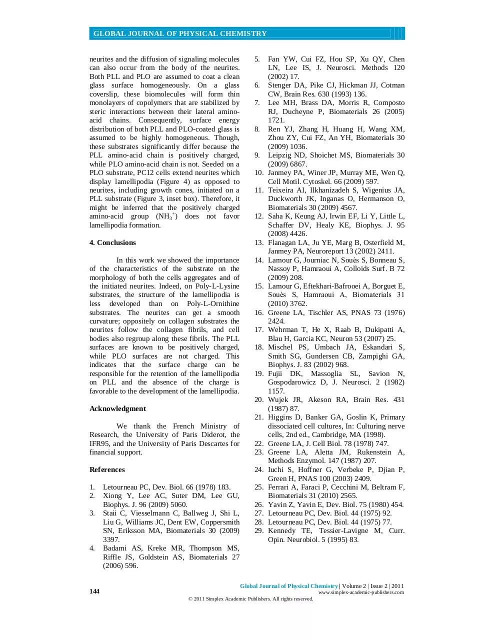
seeding) PC12 1.1 cells extend neurites whose
length may reach 100 µm (Figure 1). Though
resistant to serum deprivation, PC12 1.1 cells are
assumed to undergo some stress under these
conditions. In addition to its assumed role in
neurite initiation, this stress could also be
responsible for a reduced affinity between the
cells and the substrate. This behavior is
observable on the left image of Figure 1, where
some cells are observed exhibiting a high density
per unit area (Figure 1, arrows) which might
reflect that cells experienced poor adhesiveness
with the substrate. This lack in adhesion to the
solid surface would be compensated by cell selfclustering, which could explain the presence of
scattered patches with a high density of cells.
The arrangement of cells on the
substrate seems wholly different from what we
observed in other experiences [14, 15]. When
cells previously tended to form colonies that
appeared circular (or at least elliptical) here they
are aligned along specific directions (Figure 1).
This arrangement suggests that PC12 1.1 cells
are maintained in networks of interconnected
channels, that force cells as well as the generated
neurites to follow the directions taken by these
channels. Actually, they are most probably the
underlying collagen fibers that coat the glass
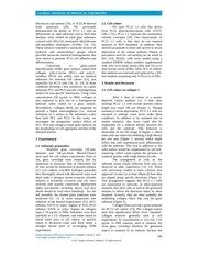
substrate (Figure 2).
Collagen fibers provide a good substrate
for PC12 cell culture [23]. The collagen matrix
used here significantly differs from “standard”
collagen matrices generally used in cell
experiments. Its concentration is very low (50
µg/mL in PBS solution, used to immerse the
clean glass coverslip) therefore the generated
matrix is assumed to be inelastic because the
Global Journal of Physical Chemistry | Volume 2 | Issue 2 | 2011
www.simplex-academic-publishers.com
© 2011 Simplex Academic Publishers. All rights reserved.
141
�
GLOBAL JOURNAL OF PHYSICAL CHEMISTRY
Figure 1. Generation of neurites by PC12 cells cultured on collagen in serum-free medium. Both cells and
neurites seem to align along specific directions. Arrows indicate cell clusters, where cells look concentrated in
high amounts. Observations were made using a phase-constrast optical microscope.
collagen layer on the clean glass is thin and
characterized by a very low roughness (rms ≤ 3.5
nm). Here, we do not discuss neither the
influence of chemical interactions between fibers
and cells, nor the impact of serum deprivation
from the culture medium on the initiation of
neurites. Rather, we note the sensitivity of PC12
1.1 cells to the nanoroughness of the glass
surface modified with collagen.
Although our previous results led us to
neglect the roughness effects on neuritogenesis
[15], it seems, at least in this work, that
nanoroughness is able to influence cell
morphology and that of the associated neurites.
Surface roughness is characterized by the rms, an
amplitude parameter which indicates vertical
deviations of the roughness profile, as well as by
morphological considerations. In other words,
surfaces sharing the same rms value can exhibit
different roughness characteristics, provided they
display diverse topographies. PC12 cells grown
on surfaces with diverse rms values (0.3 nm ≤
rms ≤ 1.4 nm) were not sensitive to rms
variations
[15],
where
topographical
heterogeneities were evenly distributed all along
the substrate. However, here the substrates
considered display specific morphological
features (i.e. the arrangement and the diameters
of collagen fibers considered at the nanometer
scale) which impact on cell and neurite growth
[25], are presumably more important than the
rms value.
3.2. Cell culture on glass-PLL and on glassPLO substrates
Figure 2. AFM images of collagen fibers adsorbed on clean glass. The root-mean-square (rms) roughness,
evaluated by the AFM software Nanoscope, is roughly equal to 3.5 nm.
142
Global Journal of Physical Chemistry | Volume 2 | Issue 2 | 2011
www.simplex-academic-publishers.com
© 2011 Simplex Academic Publishers. All rights reserved.
�
GLOBAL JOURNAL OF PHYSICAL CHEMISTRY
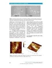
Figure 3. Generation of neurites after 6 days of culture by PC12 1.1 cells grown on glass-PLL and glass-PLO,
in a serum-deprived and NGF-free medium. The images presented here display pictures taken with a phasecontrast optical microscope, combined with an assembly of images obtained by air-tapping mode AFM, after
the cells have been fixed on the substrates using a solution of 2% glutaraldehyde in PBS.
Under the same conditions, as with
collagen matrix, PC12 1.1 cells grow neurites on
both PLL and PLO-coated glass surfaces (Figure
3) although these surfaces do not stimulate
spontaneous neuritogenesis in PC12 cells [14,
15]. It has been shown that a substrate coated
with PLL allows survival and maturation of
primary neurons in serum-free medium [26].
According to the results presented above, serum
deprivation of the culture medium is expected to
trigger neuritogenesis. Nevertheless, it is
possible that the progressive transformations
(essentially due to different kinetics of proteins
adsorption/desorption) experienced by the
PLL/PLO matrices generate local gradients in
energy of adhesion in these matrices, which
could in turn stimulate neuritogenesis [14, 15],
as well as serum deprivation. The deterioration
of the matrix created by biomolecules that do not
form covalent chemical bonds with the
underlying substrate is observed in cultures that
extend beyond three days [22, 23].
AFM imaging reveals the intense
activity of filopodia and lamellipodia in growth
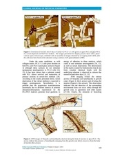
cones (Figure 4, thick arrows) and all along the
neurites (Figure 4, thin arrows). This observation
indicates that the detection of the surrounding
environment does not occur solely through the
growth cone, in agreement with other reports
[27-29]. Clearly, the initiation of branching
Figure 4. AFM images of filopodia and lamellipodia observed along the body of neurites on glass-PLO. The
arrows indicate the presence of lamellipodia emerging from the growth cone (thick arrows) or from the body
of neurites (thin arrows).
Global Journal of Physical Chemistry | Volume 2 | Issue 2 | 2011
www.simplex-academic-publishers.com
© 2011 Simplex Academic Publishers. All rights reserved.
143
�
GLOBAL JOURNAL OF PHYSICAL CHEMISTRY
neurites and the diffusion of signaling molecules
can also occur from the body of the neurites.
Both PLL and PLO are assumed to coat a clean
glass surface homogeneously. On a glass
coverslip, these biomolecules will form thin
monolayers of copolymers that are stabilized by
steric interactions between their lateral aminoacid chains. Consequently, surface energy
distribution of both PLL and PLO-coated glass is
assumed to be highly homogeneous. Though,
these substrates significantly differ because the
PLL amino-acid chain is positively charged,
while PLO amino-acid chain is not. Seeded on a
PLO substrate, PC12 cells extend neurites which
display lamellipodia (Figure 4) as opposed to
neurites, including growth cones, initiated on a
PLL substrate (Figure 3, inset box). Therefore, it
might be inferred that the positively charged
amino-acid group (NH3+) does not favor
lamellipodia formation.
5.
6.
7.
8.
9.
10.
11.
12.
4. Conclusions
13.
In this work we showed the importance
of the characteristics of the substrate on the
morphology of both the cells aggregates and of
the initiated neurites. Indeed, on Poly-L-Lysine
substrates, the structure of the lamellipodia is
less developed than on Poly-L-Ornithine
substrates. The neurites can get a smooth
curvature; oppositely on collagen substrates the
neurites follow the collagen fibrils, and cell
bodies also regroup along these fibrils. The PLL
surfaces are known to be positively charged,
while PLO surfaces are not charged. This
indicates that the surface charge can be
responsible for the retention of the lamellipodia
on PLL and the absence of the charge is
favorable to the development of the lamellipodia.
14.
15.
16.
17.
18.
19.
20.
Acknowledgment
21.
We thank the French Ministry of
Research, the University of Paris Diderot, the
IFR95, and the University of Paris Descartes for
financial support.
22.
23.
References
24.
1.
2.
25.
3.
4.
144
Letourneau PC, Dev. Biol. 66 (1978) 183.
Xiong Y, Lee AC, Suter DM, Lee GU,
Biophys. J. 96 (2009) 5060.
Staii C, Viesselmann C, Ballweg J, Shi L,
Liu G, Williams JC, Dent EW, Coppersmith
SN, Eriksson MA, Biomaterials 30 (2009)
3397.
Badami AS, Kreke MR, Thompson MS,
Riffle JS, Goldstein AS, Biomaterials 27
(2006) 596.
26.
27.
28.
29.
Fan YW, Cui FZ, Hou SP, Xu QY, Chen
LN, Lee IS, J. Neurosci. Methods 120
(2002) 17.
Stenger DA, Pike CJ, Hickman JJ, Cotman
CW, Brain Res. 630 (1993) 136.
Lee MH, Brass DA, Morris R, Composto
RJ, Ducheyne P, Biomaterials 26 (2005)
1721.
Ren YJ, Zhang H, Huang H, Wang XM,
Zhou ZY, Cui FZ, An YH, Biomaterials 30
(2009) 1036.
Leipzig ND, Shoichet MS, Biomaterials 30
(2009) 6867.
Janmey PA, Winer JP, Murray ME, Wen Q,
Cell Motil. Cytoskel. 66 (2009) 597.
Teixeira AI, Ilkhanizadeh S, Wigenius JA,
Duckworth JK, Inganas O, Hermanson O,
Biomaterials 30 (2009) 4567.
Saha K, Keung AJ, Irwin EF, Li Y, Little L,
Schaffer DV, Healy KE, Biophys. J. 95
(2008) 4426.
Flanagan LA, Ju YE, Marg B, Osterfield M,
Janmey PA, Neuroreport 13 (2002) 2411.
Lamour G, Journiac N, Souès S, Bonneau S,
Nassoy P, Hamraoui A, Colloids Surf. B 72
(2009) 208.
Lamour G, Eftekhari-Bafrooei A, Borguet E,
Souès S, Hamraoui A, Biomaterials 31
(2010) 3762.
Greene LA, Tischler AS, PNAS 73 (1976)
2424.
Wehrman T, He X, Raab B, Dukipatti A,
Blau H, Garcia KC, Neuron 53 (2007) 25.
Mischel PS, Umbach JA, Eskandari S,
Smith SG, Gundersen CB, Zampighi GA,
Biophys. J. 83 (2002) 968.
Fujii DK, Massoglia SL, Savion N,
Gospodarowicz D, J. Neurosci. 2 (1982)
1157.
Wujek JR, Akeson RA, Brain Res. 431
(1987) 87.
Higgins D, Banker GA, Goslin K, Primary
dissociated cell cultures, In: Culturing nerve
cells, 2nd ed., Cambridge, MA (1998).
Greene LA, J. Cell Biol. 78 (1978) 747.
Greene LA, Aletta JM, Rukenstein A,
Methods Enzymol. 147 (1987) 207.
Iuchi S, Hoffner G, Verbeke P, Djian P,
Green H, PNAS 100 (2003) 2409.
Ferrari A, Faraci P, Cecchini M, Beltram F,
Biomaterials 31 (2010) 2565.
Yavin Z, Yavin E, Dev. Biol. 75 (1980) 454.
Letourneau PC, Dev. Biol. 44 (1975) 92.
Letourneau PC, Dev. Biol. 44 (1975) 77.
Kennedy TE, Tessier-Lavigne M, Curr.
Opin. Neurobiol. 5 (1995) 83.
Global Journal of Physical Chemistry | Volume 2 | Issue 2 | 2011
www.simplex-academic-publishers.com
© 2011 Simplex Academic Publishers. All rights reserved.
�
 Historical archive . public document